

Aromatase inhibitors and their influence on brain – quick review
-
Poznan University of Medical Sciences

Abstract
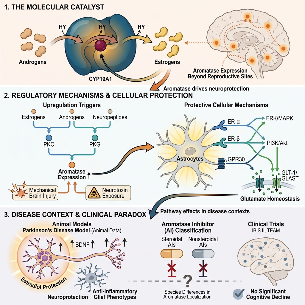
Aromatase, an enzyme responsible for converting androgens to estrogens, plays a crucial role in maintaining physiological homeostasis. It is also an essential factor in estrogen-dependent carcinogenesis. For many years, aromatase inhibitors (AIs), such as letrozole and vorozole, have been applied mainly in the treatment of breast cancer. Their utilization in managing breast and ovarian cancer, polycystic ovary syndrome, endometriosis, prostate diseases, and male infertility is well es-tablished. However, aromatase expression is not limited to classical estrogen-related tissues such as the breast, placenta, or ovaries. Its enzymatic activity is also observed in the brain, bones, and lungs. Consequently, this review focuses on the characterization of aromatase inhibitors, with particular emphasis on their adverse effects and the description of aromatase expression within the brain. The neuroprotective role of this enzyme, exhibited in response to mechanical damage and neurotoxins, is described with particular emphasis on the underlying physiological mechanisms. Furthermore, the clinical application of AIs and the role of aromatase in Parkinson's disease are also discussed.
Introduction
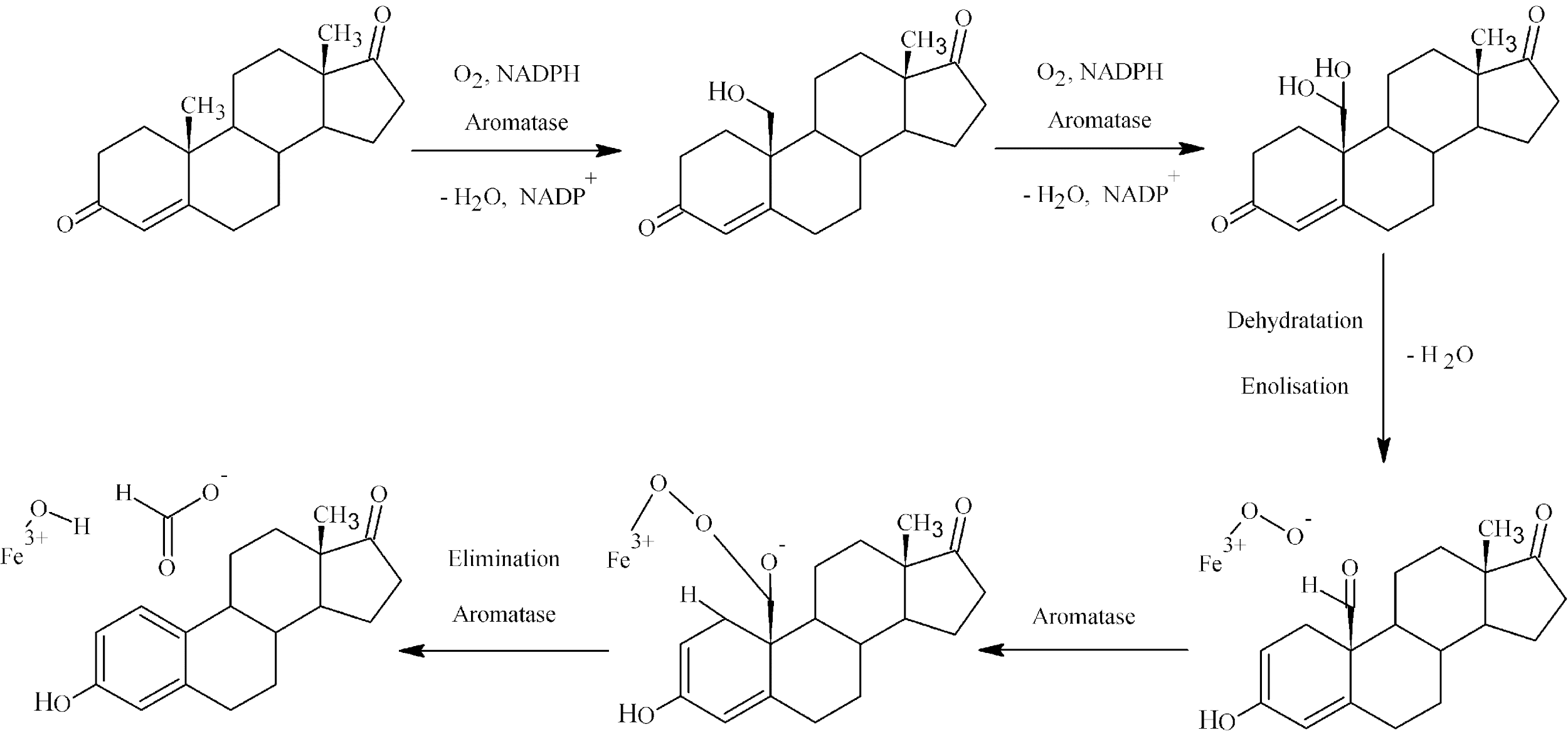
Aromatase is a metalloprotein containing a heme group as a prosthetic scaffold. This enzyme, classified as CYP19A1, belongs to the superfamily of cytochrome P450 enzymes1,2. It is responsible for converting androgens to estrogens via the aromatization of ring A of testosterone3,4 (Scheme 1).
Synthesis of estrone catalyzed by aromatase.
Aromatase is composed of two domains: α and β. The α domain is responsible for catalyzing the reaction, while the β domain is associated with substrate recognition and orientation. The heme group is sandwiched between two helices, and the region responsible for identifying the substrate is formed by four helices5. Analysis of crystallographic structures, including cocrystals with ligands, has demonstrated that the tertiary structure of aromatase consists of 12 significant α-helices and 10 β-strands dispersed across one main and three smaller sheets6. The formation of the catalytic site involves β-sheet 3, loop B-C, and helices I and F. Both polar and nonpolar amino acids participate in the key interactions between the drug and the enzyme, which are summarized in Table 1. This structural information aids scientists in designing new potential aromatase inhibitors6.
Aromatase amino acids pivotal for interacting with potential drug and properties of potential drug pivotal for interacting with aromatase Arg – arginine, Asp – aspartic acid, Ser – serine, Thr – threonine, Glu – glutamic acid, Ala – alanine, Ile – isoleucine, Leu – leucine, Val – valine, Phe – phenylalanine, Trp – tryptophan
| Aromatase amino acids crucial for binding | Potential inhibitor properties | ||
|---|---|---|---|
| Polar | Apolar | Aromatic | |
|
Arg115 Arg375 Asp309 Asp371 Ser478 Thr310 Asp371 Glu302 Met374 |
Ala306 Ala307 Ile133 Ile305 Leu477 Val369 Val370 Val373 Leu372 |
Phe134 Phe221 Trp224 |
- At least two hydrogen bonding with Met 374 - At least one hydrogen bond acceptor groups (like NO2, CN) - The capability to interact as the ligand with the iron atom of the heme group (azole – hem coordination) - Aromatic functionalized groups stabilizing the association complexes - Moieties being able to contact with the hydrophobic pocket (Ile133, Phe134, Trp224, Val370, Leu372, Val373, Met374, Leu 477) - Moieties being able to contact with the hydrophobic pocket (Ile132, Ile133, Ile305, Phe148, Met303, Ala306) |
The enzyme complex consists of two parts: aromatase and NADPH reductase6. The aromatization of the testosterone ring requires three moles of NADPH and oxygen for each mole of the obtained product, estradiol7. Overall, the reactions catalyzed by aromatase can be written as follows:
testosterone + 3NADPH + 3H + 3O ⇄ 17β-estradiol + formate + 4HO + 3NADP
androstenedione + 3NADPH + 3H + 3O ⇄ estrone + formate + 4HO + 3NADP
The reaction catalyzed by aromatase involves the aromatization of the A ring of androgens (e.g., testosterone and androstenedione) and their conversion into estrogens (e.g., 17β-estradiol and estrone). The mechanism occurs according to the scheme below and generally involves three successive hydroxylations (Scheme 1).
Aromatase Inhibitors (AIs)
Aromatase inhibitors (AIs) inhibit aromatase activity and reduce estrogen concentrations. These drugs can be classified into three generations, as well as into two structural groups: steroidal and nonsteroidal8 (Figure 1). The first generation of these drugs originally had alternative clinical applications prior to being identified as aromatase inhibitors; for example, aminoglutethimide was applied as an antiepileptic drug, and testolactone exhibited androgenic activity8. However, their limited specificity and numerous adverse effects encouraged the development of new derivatives.
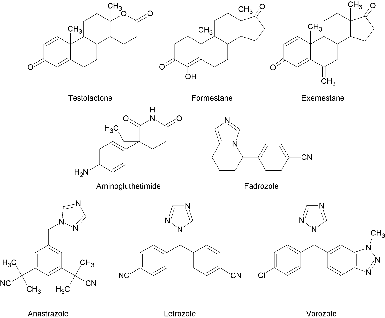
Classification and chemical structures of steroidal and nonsteroidal aromatase inhibitors. Aromatase inhibitors (AIs) are categorized into two main structural groups across three generations of drug development. Steroidal AIs (e.g., testolactone, formestane, and exemestane) are analogs of natural hormones that imitate the natural substrate to irreversibly block the enzyme. Nonsteroidal AIs (e.g., aminoglutethimide, fadrozole, anastrozole, vorozole, and letrozole) represent a heterogeneous group of compounds that act through reversible enzyme inhibition.
Steroidal AIs are analogs of natural hormones, mainly androstenedione, with testolactone being the first drug classified in this group. Initially, it was supposed that its mechanism of action was strongly androgenic, but subsequent investigations demonstrated that it inhibits aromatase9. Further research identified additional inhibitors, such as formestane and exemestane (Figure 1). Generally, the action of steroidal AIs is based on imitating the natural substrate by fitting into the active center of the enzyme. Subsequently, aromatase transforms the drug into a reactive intermediate, which irreversibly blocks the enzyme9.
Nonsteroidal AIs comprise a heterogeneous group of medicaments, with aminoglutethimide being the first discovered and classified in this group. It was originally applied as an antiepileptic drug, but due to adverse effects (mainly insufficiency and ataxia) and poor efficacy, it was withdrawn from treatment. Furthermore, clinical trials showed that aminoglutethimide inhibits both aromatase and cytochrome P450scc. The latter enzyme catalyzes the conversion of cholesterol to pregnenolone9. The unintended inhibition of P450scc prompted scientists to search for more selective AIs. This research led to the discovery of fadrozole, anastrozole, vorozole, and letrozole (Figure 1)9.
The mechanism of action of nonsteroidal AIs, described herein using the example of letrozole, is based on reversible enzyme inhibition. These compounds fit into the active site and create a strong coordination bond with iron (II) within the heme group. The moiety capable of forming hydrogen bonds with the enzyme is crucial for enzyme inhibition (e.g., a cyano moiety). In addition, the structure is stabilized by a π-electron system, and the nitrile group allows for good fitting within the β domain of the enzyme10. This interaction is explained by analogy to the carbonyl group of androstenedione, the natural substrate11.
Figure 2 shows the docking of androstenedione and letrozole into human aromatase. The cyan color indicates androstenedione and the pink color indicates letrozole; interactions with the heme group (purple) and hydrogen bonding interactions with amino acids Arg115, Met374, or Ser478 are visible.
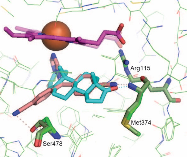
The docking of letrozole and androstenedione into the human aromatase. Letrozole (marked in pink) is in the light green protein and androstendione (marked in cyan) is in the dark green protein. The haeme group is in purple with the iron shown as orange sphere and possible hydrogen bonds are shown by dotted lines. Modified according to
Side Effects
Aromatase inhibitors (AIs) constitute an important class of drugs used in the treatment of hormone-dependent breast and ovarian cancer in postmenopausal women12. The application of third-generation drugs, which exhibit high affinity for aromatase, contributes to their overall safety profile. They cause a limited number of adverse effects, primarily hot flashes, joint pain, weight gain, fatigue, insomnia, mood swings, vaginal discharge, and vaginal dryness 13,14.
These adverse effects are associated with a decrease in estrogen concentration. Another known complication is an increased risk of osteoporosis. When estrogen is at physiological levels, it acts via the activation of ER-α receptors in osteoclasts. This leads to the activation of the Fas/FasL pathway, apoptosis induction, and the inhibition of bone resorption15. In the absence of estradiol, the synthesis of pro-inflammatory cytokines — including IL-1, IL-6, TNF-α, granulocyte-macrophage colony-stimulating factor (GM-CSF), macrophage colony-stimulating factor (M-CSF), and prostaglandin E2 (PGE2) — occurs16. Normally, estradiol inhibits the activity of IL-1 and TNF-α, which are potent stimulants of bone resorption. Their secretion is mediated by monocytes circulating in the blood and is increased in postmenopausal women and during osteoporosis. Additionally, both cytokines induce the production of IL-6, which stimulates the proliferation of early hematopoietic cells that differentiate into osteoclasts.
Estradiol can inhibit the induction of IL-616. Furthermore, estradiol increases the synthesis of procollagen, IGF-1, and TGF-β in osteoblasts and extends the lifespan of osteoblasts by inhibiting apoptosis. The increase in TGF-β synthesis blocks bone loss by inhibiting osteoclast activity and promoting their apoptosis16.
The osteoblastic stromal cell line can express RANKL (receptor activator of NF-κB ligand), which causes the activation of RANK (receptor activator of NF-κB) on the surface of osteoclasts. Although the activation of RANK leads to osteoclast maturation, this process alone is insufficient, as a suitable concentration of M-CSF is also necessary. In addition to RANKL, osteoblast stromal cells secrete osteoprotegerin (OPG), a decoy receptor for RANKL. OPG binds to RANKL, deactivating it and thereby inhibiting osteoclast development and apoptosis. The cytokines IL-1 and TNF-α cause an increased production of RANKL, M-CSF, and OPG. PGE2 reduces the synthesis of OPG and increases RANKL. In turn, estradiol production increases OPG and reduces the formation of M-CSF and RANK16.
Similarly, androgens exert an impact on osteoblast growth, proliferation, and differentiation. In contrast, this results in a lower production of OPG. 5-α-dihydrotestosterone, a potent androgen, increases the synthesis of IGF-1, IGF-II, and TGF-β mRNA, and decreases the synthesis of IL-6 16,17. A study on male rats demonstrated that protection from bone loss is mediated by AR and ER receptors. In the absence of estradiol or a lack of aromatase, a loss of bone mass was noticed. This effect was not observed in females. However, in females, the physiological level of estrogen is higher, which may partly explain these experimental results. AR receptors may exert a protective effect only when estrogen levels decrease, e.g., during menopause or during pharmacotherapy with AIs18.
Selective androgen and estrogen receptor modulators activate ARs in a tissue-specific manner, providing a safe alternative in the treatment of osteoporosis and muscle disease19. For example, the compound S-040503 increased bone mineral density and the biomechanical strength of the femoral cortex in a rat model of orchidectomy19. Similar results were obtained in animal models of postmenopausal osteoporosis using ovariectomized rats19. However, further studies on the modulation of AR signaling and its effects on bone structure and metabolism are necessary19.
According to literature data, the development of osteoporosis can also be affected by other factors: calcium and vitamin D deficiencies20; the use of certain medicaments (e.g., phenytoin21, barbiturates21, and glucocorticosteroids22); and concomitant diseases (e.g., diabetes and Cushing's syndrome). Nevertheless, hormones play a vital role in protecting against bone diseases22.
Although steroidal and nonsteroidal AIs produce the same therapeutic effect (lowering estrogen levels), it appears that the risk of osteoporosis is more closely associated with nonsteroidal agents23. Studies in rats have shown that exemestane and its metabolite—17-hydroexemestane—can inhibit bone loss and increase bone strength23. This effect is explained by the androgenic activity of the drug and its metabolite23. Furthermore, treatment with exemestane has been shown to reduce the incidence of bone pain, fractures, and osteoporosis compared with anastrozole and letrozole24. A smaller decline in bone mineral density was observed compared with anastrozole24. Overall, exemestane reduced the incidence of bone-related adverse events24.
However, a two-year human study demonstrated that exemestane may affect the development of osteoporosis25. In some patients with pre-existing osteopenia, the condition progressed to osteoporosis. In patients without initial osteopenia, osteoporosis did not develop25. Physiological differences between rats and humans, as well as differences in exemestane dosages, may explain the discrepancies between these studies25. However, human studies have shown that in addition to an increase in bone resorption markers (e.g., CTX - C-telopeptide), there is also an increase in bone formation markers (e.g., BAP - bone alkaline phosphatase)25.
In discussions regarding the adverse effects of AIs, much attention is paid to the risk of osteoporosis or arthralgia, while little attention is given to their effects on brain function. Estradiol affects mood and cognitive function, and it exhibits a preventive action against Alzheimer's disease26,27,28. However, current studies have not definitively answered the question of the impact of AIs on cognitive function29. Only one conclusion is indisputable: future studies should be conducted on large populations.
Cognitive impairment may be related to low circulating levels of estradiol in patients. There are at least two randomized trials that were statistically powered to detect moderate (but not small) effects, representing an important caveat. The studies themselves have not proven that aromatase inhibitors adversely affect cognition or that they have a greater effect than tamoxifen29. No cognitive decline was observed in a cohort study of women taking aromatase inhibitors for one year (assessed using the Mini-Mental State Examination - MMSE). However, this study showed significant (p < 0.05) changes after 6 and 12 months of treatment in terms of depressive symptoms (measured by the Geriatric Depression Scale) and sleep quality (measured by the Athens Insomnia Scale - AIS)30.
The effects of letrozole on cognitive function, anxiety, thermoregulation, brain estrogen content, and hippocampal pyramidal cell physiology are noteworthy. Estradiol levels in the hippocampus were shown to increase in both females and males. This was likely the cause of the negative effect on spatial working memory and the intrinsic excitability of hippocampal neurons. Despite a lack of changes in hypothalamic estradiol levels, thermoregulatory disorders were observed in females. This may translate into the observed adverse effects in humans31.
Third-generation AIs exhibit high specificity, but apart from the systemic inhibition of aromatase, primarily in cancer tissues, they also inhibit aromatase in the brain. Among the available nonsteroidal AIs, anastrozole reaches the lowest concentration in the brain, while letrozole and vorozole achieve relatively high concentrations32. The low concentration of anastrozole in the brain is caused by the activity of P-glycoprotein (an efflux pump responsible for the active removal of drugs from cells, which is also associated with resistance to cancer treatment). Letrozole, in turn, is a poor substrate for this transporter, and this fact has no clinical significance. Although vorozole is a substrate for P-glycoprotein, it reaches high concentrations in the brain, which is attributed to its excellent lipid permeability32,33. Considering these factors, possible neurological adverse effects related to AI treatment should be carefully investigated.
The Expression of Aromatase in the Brain
Aromatase is widely distributed throughout the body. Beyond its primary sites of expression—such as the gonads, breast, and placenta—it is also found in adipose tissue, muscle, bone, and the brain34,35.
The coding region of the aromatase gene includes nine exons, starting from exon II. Preceding this exon is exon I, which is regulated by alternative tissue-specific promoters in the 5'-untranslated region. Although each tissue expresses aromatase using a tissue-specific transcriptional promoter linked to exon I, the resulting translation product is always identical; therefore, the aromatase protein structure is the same across all tissues34,35.
In the brain, aromatase expression is upregulated by several factors, including estrogens, androgens, substance P, cholecystokinin, neurotensin, brain natriuretic peptide, dibutyryl cGMP, and α1-adrenergic agonists. The involvement of these substances indicates that the leading regulatory pathways are mediated by protein kinase C (PKC) and protein kinase G (PKG). In contrast, cyclic AMP (cAMP) signaling can suppress or otherwise modulate aromatase expression (Table 2)36.
Selected neurotransmitters and mechanisms involved in expression of aromatase.
| Neurotransmitters | Mechanism |
|---|---|
| noradrenaline, adrenaline | cAMP |
| protein kinase A | |
| noradrenaline, adrenaline | diacylglycerol and inositol 1,4,5-trisphosphate |
| protein kinase C | |
| substance P | diacylglycerol and inositol 1,4,5-trisphosphate |
| protein kinase C | |
| cholecystokinine | diacylglycerol and inositol 1,4,5-trisphosphate |
| protein kinase C | |
| neurotensin | diacylglycerol and inositol 1,4,5-trisphosphate |
| protein kinase C | |
| natriuretic peptides | cyclic guanosine monophosphate |
| protein kinase G |
Neuroprotection Against Damage
Aromatase plays a variety of functions in the brain, with one of the most important being neuroprotection37,38,39,40. When brain damage occurs (whether mechanical or toxin-induced), an increase in aromatase expression and activity is noted37,38,39. This phenomenon is observed in glial cells, where normal expression is typically low, while in neurons, it is maintained at a physiological level at all times37,38,39. This upregulation occurs in both sexes and across all damaged areas of the brain, including the hippocampus, cortex, corpus callosum, striatum, and hypothalamus40. Additionally, aromatase protects the nigrostriatal system and substantia nigra39,40. Further studies in animal models should be conducted to better understand the pathways of aromatase expression during ischemia41,42.
Neuronal protection in the hippocampus and inferior olivary nucleus has been demonstrated in studies utilizing region-specific neurotoxins (e.g., kainate for the hippocampus and 3-acetylpyridine for the inferior olive). Androgen receptor knockout (ArKO) animals and those receiving AIs exhibited more extensive brain damage than animals with undisturbed aromatase function39,40,43. Another study confirmed the neuroprotective function of extragonadal aromatase for hippocampal neurons, highlighting their increased susceptibility to excitotoxic damage when aromatase is inhibited. It was demonstrated that neurons in both male and female rats receiving fadrozole, as well as in ArKO models, were more susceptible to injury39,40.
The effect of testosterone and estradiol on the neuroprotection activity of aromatase was also investigated. Studies utilizing formestane and flutamide (an anti-androgen) have shown that testosterone exerts a protective effect on neurons43. The neuroprotective effect of testosterone was comparable both in the presence and absence of AIs; however, in the presence of flutamide, these protective effects were inhibited43. However, another study showed that co-administering additional neurotoxins, e.g., kainate, with testosterone diminished this protective function. In one experiment, male rats were treated with testosterone in the presence of fadrozole. The addition of the AI resulted in greater damage to neural connections, indicating a significant role for aromatase in brain protection43.
The mechanism of aromatase's neuroprotective effect is multifactorial, involving the synthesis of estradiol, activation of nuclear and membrane receptors, estrogen receptor interaction with IGF-1R (insulin-like growth factor 1 receptor), antioxidant activity, and non-receptor interactions with other cells. Estradiol mediates neuroprotective actions through both neurons and astrocytes. Various studies have attempted to assess the relative contributions of these two cell types and ultimately concluded that astrocytes play a predominant role in neuroprotection, whereas the role of neurons is secondary44,45.
Dying neurons release cations and glutamate into the extracellular space. This leads to the activation of microglia, which subsequently produce the cytokines IL-1 and IL-6. Astrocytes express a large variety of surface receptors, including all subtypes of estradiol receptors (the classical ER-α and ER-β, G protein-coupled ER, and membrane GPR30 receptors), EGF receptors, IGF-1 receptors, TGF-β receptors, cytokine receptors, TNF receptors, glutamate receptors, and ion channels46,47. Cytokines and glutamate can modulate inflammation and neuronal damage47. The effect of cytokines on aromatase activity remains under investigation. Zwain et al. demonstrated that interleukin (IL)-1β induces a dose-dependent inhibition of aromatase48. Conversely, other research indicates that IL-1β and IL-6 expression is associated with increased aromatase activity49. These divergent results indicate that further studies are needed to elucidate the effect of cytokines on aromatase expression. However, one definitive conclusion can be drawn: the impact of cytokines depends on the regional origin of the astrocytes.
Astrocytes express two primary glutamate transporters on their surface: the GLAST receptor (also known as EAAT1, excitatory amino acid transporter 1) and the GLT-1 receptor (or EAAT2, excitatory amino acid transporter 2)39,40. Glutamate can activate a variety of receptors, including the ionotropic NMDA, AMPA, and kainate receptors, as well as metabotropic receptors (e.g., mGluR). An excessive increase in the activity of NMDA and AMPA receptors can lead to excitotoxicity through ionic imbalance, specifically an influx of calcium and sodium ions into the neuronal cell50,51. The presence of GLAST and GLT-1 facilitates glutamate uptake from the synaptic space and prevents excitotoxicity51,52. When a brain injury occurs, the expression of these transporters reduces, which results in an expansion of the damaged area.
Estradiol affects the expression of these transporters by activating ER-α and ER-β receptors to modulate GLT-1 expression. While the mechanism of regulation by ER-α and ER-β receptors is not fully understood, GPR30 receptor stimulation activates several pathways, including ERK/MAPK, PI3K/Akt, PKA, and Src47. The control of GLT-1 is also mediated by NF-κB (a transcription factor protein complex) and TGF-α (transforming growth factor-α). Estradiol, via the GPR30 receptor, induces the binding of the p50 and p65 subunits of NF-κB to the GLT-1 promoter47. It also causes CREB phosphorylation, an increase in CRE activity, and the induction of CREB binding to the GLT-1 promoter.
The impact of TGF-α is driven by an increase in its synthesis and release following the activation of GPR30, ER-α, and ER-β receptors. The released TGF-α acts via an autocrine mechanism on the EGF receptor, and its activation stimulates intracellular pathways that lead to an increase in GLT-1 expression. In addition, estradiol causes the phosphorylation of the EGF receptor, which results in increased activity of both TGF-α and EGFR.
The mechanism of increased GLAST expression is different from that of GLT-1 because nuclear ER-α and ER-β receptors do not directly affect it53. Instead, by activating its membrane receptors, estradiol causes the activation of the PI3K/Akt pathway, which is responsible for the increased expression of TGF-β (transforming growth factor-β, belonging to the transforming growth factor β superfamily)54. TGF-β, either occurring intracellularly or activating the TGF receptor on the cell surface, results in the activation of the PI3K/Akt and MAPK/ERK pathways, which leads to an increase in GLAST expression. The same mechanism occurs through other growth factors, such as IGF-1 or bFGF (basic fibroblast growth factor). PKC (protein kinase C) is another factor involved in the regulation of GLAST.
The presence of ER-α and ER-β receptors on the surface of astrocytes is necessary for their neuroprotective activity55,56. Activation of ER-α leads to an anti-inflammatory effect, which is mediated through the inhibition of various inflammatory factors. Activation of the nuclear estrogen receptor suppresses the transcriptional activity of inflammatory factors dependent on NF-κB activation by inhibiting the attachment of the p65 subunit to the chemokine CCL2 promoter57. Additionally, interfering with NF-κB and AP-1 inhibits the formation of MMP-9 (metalloproteinase) mRNA56.
The regulation of GLAST/GLT1 transporters by estradiol seems particularly intriguing and may occur, for example, via: a) PI3 kinase coupled with NO production58, b) transcriptional control via CREB59, or c) nuclear estrogen receptors60. Several factors that modulate the expression and function of GLAST/EAAT1 and GLT-1/EAAT2 are listed in Table 3, but the topic itself requires further exploration61,62.
Factors modulating the expression and function of GLAST/EAAT1 and GLT-1/ EAAT2. Modified according to
| Factor | Signaling path |
|---|---|
| β-lactam antibiotics | PI3K-Akt, NF-κB |
| tamoxifen | PI3K-Akt, TGF-α, ERK, NF-κB and nuclear ER binding |
| brain-derived neurotrophic factor | BDNF-TrkB, CREB, NF-κB |
| basic fibroblast growth factor | MAPK ERK, NF-κB, PI3K-Akt |
| valproic acid | Class I and HDAC inhibition, histone acetylation |
Estradiol also exhibits anti-inflammatory activity by reducing the expression of cyclooxygenase-2 and inducible nitric oxide synthase, and increasing the expression of the PPAR receptor (peroxisome proliferator-activated receptor). PPAR receptor activation is characteristic of nonsteroidal anti-inflammatory drugs. It suppresses the inflammatory protein Aβ (β-amyloid), which is responsible for neuroinflammation and neuronal destruction in Alzheimer's disease. Estradiol does not directly activate the receptor but enhances its expression and induces its activation63. The PPAR receptor activator is formed by coupling estradiol with the metabolism of arachidonic acid64. Moreover, receptor activation leads to the suppression of NF-κB and promotes anti-inflammatory actions63.
Sex-dependent differences in astrocyte sensitivity to harmful factors and estradiol activity have been documented. Male astrocytes are more sensitive to oxidative stress, oxygen and glucose deprivation, toxins, organophosphates (e.g., DMT - Dimethoate), and LPS (Lipopolysaccharide) compared to female astrocytes65,66,67,68. In males, LPS exposure results in a greater increase in IL-1β, IL-6, and TNF-α than in females66. The same applies to dimethoate (DMT) exposure, with the difference that DMT generally does not increase pro-inflammatory cytokines in female astrocytes67.
Studies have examined estradiol's impact on activating individual ER-α and ER-β receptors and their ability to inhibit pro-inflammatory factors. In one study utilizing female astrocytes, the effect of LPS on the induction of inflammation was investigated. Estradiol, through the ER-α receptor, inhibits only the formation of IL-1β, whereas through the ER-β receptor, it inhibits the formation of IL-1β, TNF-α, and MMP-968. In another study using DMT on male and female astrocytes, it was demonstrated that activation of the ER-α receptor induced the downregulation of IL-1β. In contrast, activation of ER-β caused a decrease in IL-1β, IL-6, TNF-α, and IP10. This effect was observed in male astrocytes but not in female astrocytes. These differences may result from inherent sex-based variations in inflammatory responses67. Additionally, ER-α activation inhibits lymphocyte and macrophage responses69.
Although astrocytes play a primary role in neuroprotection, the role of neurons must also be acknowledged. In neurons during injury, aromatase activity increases, leading to an increase in estradiol levels. Neurons also express all the types of estrogen receptors mentioned previously. Their activation leads to different intracellular signal transduction pathways compared to astrocytes. In neurons, activation of ER-α leads to an increased opening of BK (Big Potassium) ion channels, leading to increased cellular hyperpolarization, which blocks the excitatory pulse from excessive NMDA glutamate receptor activation70. Additionally, the activation of nuclear ER-α and ER-β receptors increases the expression of Bcl-2 (an anti-apoptotic protein)71.
The individual role of ER-β in neuronal neuroprotection is primarily localized to the mitochondria. Its activation can inhibit apoptotic pathways, and the receptor regulates the activity of mtDNA and complex IV of the electron transport chain72. Not only can ER-α and ER-β receptors be found in neurons, but also membrane estrogen receptors, especially the GPR30 receptor, which play essential functions in regulating neuronal survival and apoptosis. Through activation of this receptor by estradiol, ERK1/2 (a complex of proteins that transmit information from the cell membrane) is activated. Activation of this pathway leads to the subsequent activation of Akt, also known as Protein Kinase B73.
Akt belongs to the serine-threonine protein kinase family, enzymes responsible for the phosphorylation of proteins, mainly at serine or threonine residues. Phosphorylation changes protein conformation, leading to their activation or deactivation. Akt activation leads to CREB activation73; this cellular transcription factor, upon binding to a suitable DNA fragment, promotes the transcription of pro-survival genes. In the case of estradiol, this leads to the transcription of BDNF (brain-derived neurotrophic factor), which is necessary for neuronal function. Activation of ERK1/2 also leads to the inhibition of p-DAPK-1 dephosphorylation, a kinase associated with apoptotic activities. By inhibiting dephosphorylation, the active form of DAPK-1 is not formed, leading to the phosphorylation of the NMDA glutamate receptor (especially the NR2B subunit)74. The result is decreased activity of the NMDA receptor, which protects against excitotoxicity74.
Estradiol, via GPR30 receptor activation, similarly to ER-α and ER-β, leads to increased Bcl-2 expression; however, activating the membrane receptor also inhibits the degradation of procaspase-3. Preventing the activation of caspase-3 halts apoptotic pathways75. Another effect of estradiol binding to the GPR30 receptor is the inhibition of JNK, a kinase which otherwise leads to the activation of apoptotic pathways. Figure 3 and Figure 4 briefly present the activities of estradiol leading to neuroprotection and the inhibition of inflammation and reactive gliosis. Figure 5 presents a more detailed connection between estrogen, tamoxifen, and neuroprotective activity.
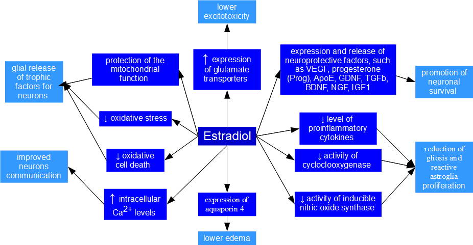
Selected activities of estradiol leading to neuroprotection. The schematic illustrates key intracellular signaling pathways activated by estradiol to promote neuronal survival. These mechanisms include: (1) the activation of the ERK1/2 and Akt signaling cascades, leading to CREB-mediated transcription of brain-derived neurotrophic factor (BDNF); (2) the inhibition of p-DAPK-1 dephosphorylation via ERK1/2, which decreases NMDA receptor activity to prevent excitotoxicity; and (3) the suppression of apoptotic pathways through the modulation of Bcl-2, inhibition of JNK, and prevention of caspase-3 activation. Glutamate receptors (NMDA and AMPA), as well as calcium and sodium ions, are highlighted in red.
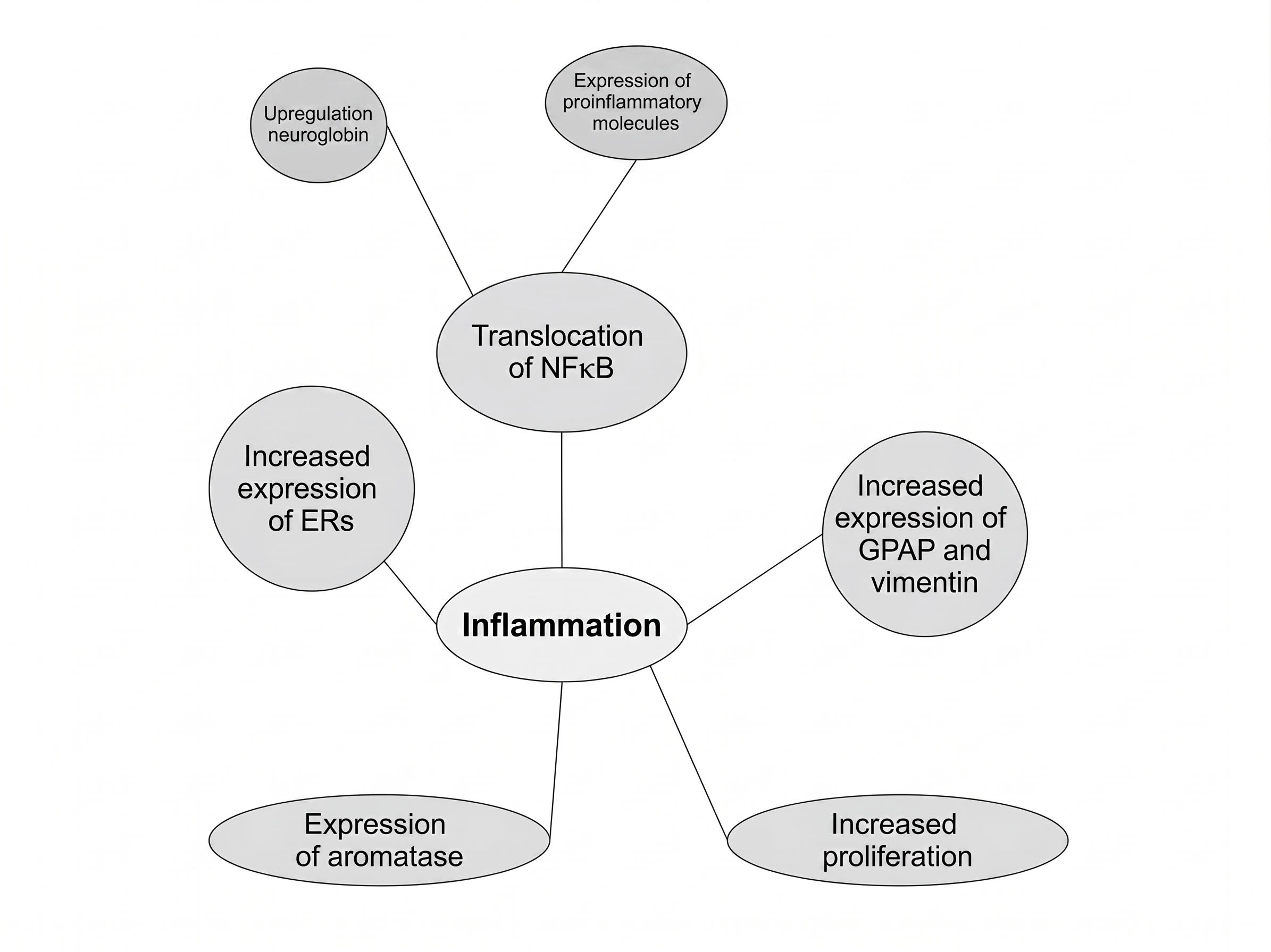
Selected activities of estradiol leading to the inhibition of inflammation and reactive gliosis. The schematic illustrates the anti-inflammatory mechanisms of estradiol within the brain. Estradiol exerts its protective effects by downregulating the expression of key pro-inflammatory enzymes, specifically cyclooxygenase-2 (COX-2) and inducible nitric oxide synthase (iNOS), thereby preventing inflammation-induced neuronal destruction. Additionally, estradiol promotes anti-inflammatory signaling by coupling with arachidonic acid metabolism to generate ligands that activate peroxisome proliferator-activated receptors (PPAR). These actions collectively suppress neuroinflammation and limit excessive reactive gliosis. Prostaglandin E2 (PGE2) is highlighted in red.
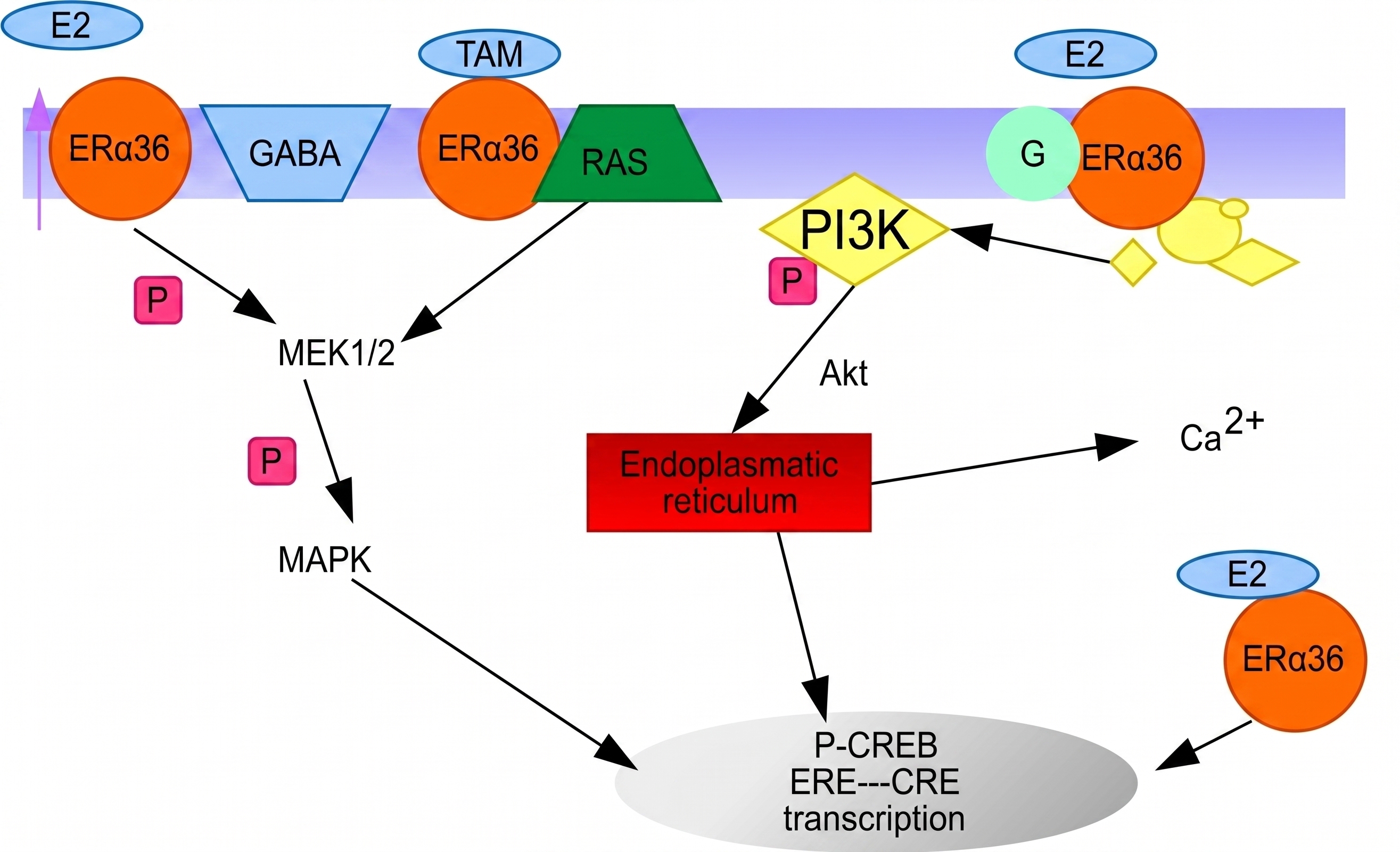
Neuroprotective action of estrogen (E2) and tamoxifen (TAM). Prepared based on
Undoubtedly, the broadly discussed inflammatory conditions are strongly associated with neuroprotective mechanisms. Understanding all the factors and signaling pathways that connect these conditions requires further intensive research. In the literature, there are reports on: a) nicotinic acetylcholine receptors, which activate the main regulator of oxidative stress, Nrf2/HO-1, via the cholinergic anti-inflammatory pathway (inhibition of TNF-α release induced by LPS, cyclooxygenase-2, and prostaglandin E2 synthesis; reduced release of TNF-α and IL-1β; and activation of ERK1/2 and p38) and the anti-inflammatory pathway associated with the activation of the Jak2/STAT3 pathway (inhibition of NF-κB nuclear translocation)76; b) enolase - its increased levels lead to the increased production of pro-inflammatory cytokines, chemokines, and some growth factors. Furthermore, NSE (neuron-specific enolase) may also have a neurotrophic function, as it controls neuronal survival, differentiation, and the regeneration of neural processes by activating the PI3K/AKT and MAPK/ERK pathways77. Moreover, NF-κB plays a role in regulating the pathogenesis of neuroinflammation-related diseases. An interesting study on NF-κB dimers demonstrated their equivocal ability to inhibit both proapoptotic and antiapoptotic pathways78.
The key clinical importance of aromatase in astrocytes stems from the fact that these cells are its main source in the brain after injury and during inflammatory conditions. From a medical perspective, it is important to note that astrocytes in both sexes increase aromatase production in response to brain damage (e.g., stroke or mechanical trauma)79,80. However, in females, this mechanism often provides stronger neuroprotection, as locally produced estradiol inhibits excessive astrocyte reactivity and protects neurons from excitotoxic death81. Furthermore, male astrocytes exhibit distinct dynamics of aromatase expression82,83. The lack of a strong estrogen response comparable to that in females may make male brains more susceptible to chronic inflammation following neurological events.
It is also important to emphasize that aromatase-blocking drugs are used in oncological treatments (breast cancer). This has direct clinical implications for cognitive function in women taking AIs; blocking aromatase in astrocytes can lead to so-called aromatase-induced atrophy, "brain fog", or an increased risk of depression and neurodegenerative diseases, as the brain loses its natural estrogenic protection84,85,86. Finally, aromatase efficiency in astrocytes appears to decline with age. In postmenopausal women, this decline is more dramatic, which, combined with sex differences in astrocyte responses, may explain the higher incidence of Alzheimer's disease87,88.
In conclusion, astrocyte aromatase acts as an "internal rescue system." Sex differences in its efficiency determine how effectively a patient's brain can autonomously limit damage. In summarizing the adverse effects and neuroprotective functions associated with aromatase, estrogens, and AIs, Table 4 may be useful. Common elements of biochemical pathways and neurotransmitters are marked in red. Further research would be worthwhile to deepen our knowledge of the interrelationships between IGF1, cytokines, and NF-κB.
Summary of key signaling pathways involved in osteoporosis and neuroprotection. Common mediators are highlighted in red.
| Mechanism influencing by and related to osteoporosis | Mechanism related to neuroprotective role of aromatase and estradiol | |||
|---|---|---|---|---|
| Osteoclast | Osteoblastic stromal cell line | Osteoblast | Neurons | Astrocytes |
| Fas/FasL pathway | RANK/RANKL | OPG | ERα: BK potassium ionic channel |
ERα/β receptors - TGFα |
| IL-1, Il-6 | NF-κB | IGF-1 | ERα/β receptors: Bcl-2 |
GPR30 receptor: - ERK / MAPk - PI3K / Akt - PKA - Src - TGF-α |
| TNFα | OPG | IGF-II | ERα: mitochondial activity |
EGF receptor - TGFα - EGFR |
| GM-CSF and M-CSF | IL-1 | TGF-β |
GPR30 receptor: - ERK1/2 activation and Akt activation, (CREB activation and transcription of BDNF) - ERK1/2 activation and p-DAPK-1 dephosporylation (NMDA lower activity) - Bcl-2 and caspase-3 activity - inhibition of JNK | Glutamate receptors: NMDA receptors, AMPA receptors, calcium and natrium ions |
| PGE2 | TNFα | IL-6 |
GLT-1 receptor - TGF-α | |
| M-CSF | Cytokines: IL-1B and IL-6 | |||
| PGE2 | Expresion of COX-2, iNOS, PPAR receptor | |||
| IGF1 receptors | ||||
A summary of the various research effects on the neuroprotective role of aromatase and the influence of aromatase inhibitors on cognitive functions is presented in Table 5. It summarizes the key conclusions from the research and provides additional comments on further research directions. The clinical controversy regarding the effects of aromatase inhibitors on cognitive function stems from discrepancies between animal and human studies. While animal models consistently demonstrate a negative effect of aromatase inhibitors on memory and brain plasticity, clinical data in breast cancer patients remain ambiguous and often contradictory87,88,89,90,91,92,93,94,95,96,97,98,99,100,101,102,103,104,105,106,107,108. This controversy stems primarily from a discrepancy between animal models and human studies. For example, in rodents and birds, aromatase inhibition almost always leads to marked deficits in spatial and working memory, as well as impaired synaptic plasticity. Studies in knockout mice (ArKO) confirm that the complete absence of this enzyme disrupts the development and functioning of neuronal circuits99,100,101,102.
Summary of studies on the effects of aromatase inhibitors on cognitive functions. Based on
| Assay | Compared drugs | Outcomes | Analysis and Controveries |
|---|---|---|---|
| BIG 1-98 | Letrozole | Patients taking letrozole had better overall outcomes than those on tamoxifen |
It has been suggested that tamoxifen, rather than aromatase inhibitors, has a more pronounced negative effect on the brain. This is due to tamoxifen's ability to modulate the estrogen receptor, which may antagonize key cognitive domains. Cognitive function appears to improve significantly one year after discontinuing hormonal therapy (both AI and tamoxifen), suggesting that the effects of the drugs on the brain are at least partially reversible. |
| IBIS II | Anastrozole | No significant differences in cognitive function between the treatment and placebo groups | The demonstrated lack of effect on cognitive function contradicts the subjective complaints of the patients and molecular biology data. Perhaps more sensitive cognitive tests should have been used. |
| TEAM | Exemsetane | No evidence of moderate or major negative effects of AI compared with tamoxifen | The placebo effect cannot be assessed because the study groups were taking medication. It is possible that exemestane also binds to androgen receptors. Therefore, there is a risk that tamoxifen may mask the negative effects of estrogen deprivation. |
| ATAC | Anastrozole | It showed poorer learning and memory scores (visual and verbal) in women taking anastrozole compared to tamoxifen. | The statistical significance of the test results and their clinical sensitivity are controversial. Furthermore, patients taking both medications did not perform better in the tests. This suggests that aromatase inhibitor-induced astrocyte damage is irreversible. The statistical strength of these conclusions is questionable due to the representativeness of the sample and the age of the patients. |
| García-Sánchez et al. (2022) | Anastrazole and letrozole | Adjuvant aromatase inhibitor (AI) therapy significantly worsens mood and sleep quality in post-menopausal women. | Higher sensitivity methods were used. The focus was on a relatively small study sample and no control group. |
| Phillips et al (2011) | - | Systematic analysis of large clinical trials and comparison of results with those obtained in animal models. |
Based on molecular mechanisms, one might expect that drastic reductions in estrogen levels, which are crucial for synaptic plasticity, would lead to severe cognitive deficits. However, data from the IBIS-II and TEAM studies do not support the existence of moderate or large negative effects of aromatase inhibitors on cognitive function compared with placebo or tamoxifen. It is necessary to select tests with appropriate sensitivity. Publications indicate that large studies were sufficiently powered to detect moderate changes; however, smaller deficits cannot be ruled out, which may be bothersome for an individual patient (e.g., a professionally active person). Perhaps the tests used in these studies are too simple to detect subtle changes in the hippocampus, which are evident in animal models. Longer-term studies are needed. It's possible that 5 years (or longer) of aromatase blockade in astrocytes doesn't accumulate damage that becomes apparent only after treatment ends, increasing the risk of Alzheimer's disease in later life, for example. There is a need to appropriately select the study groups of patients taking into account the chemotherapy they have previously received. |
| Yuste et al (2025) | - | Identification of potential pharmaco-vigilance signals related to dementia and Alzheimer's disease and third-generation aromatase inhibitors in menopausal and post-menopausal women. | A disproportionate reporting of dementia, Alzheimer's disease, and senile dementia has been demonstrated with aromatase inhibitors. This is consistent with knowledge of estrogen and aromatase function and preclinical studies. However, the publication only covers adverse event reports, which already poses a significant risk to humans. |
| Anastrozole, exemestane, or letrozole | No harmful effects of aromatase inhibitors on cognitive function were observed in post-menopausal breast cancer patients. | A causal relationship between hypertension and cognitive decline was demonstrated. | |
| Branigan et al (2020) | Tamoxifen, exemestan, other aromatase inhoibitors | The use of tamoxifen and steroid aromatase inhibitors was associated with a reduction in diagnosed neurodegene-rative diseases (including Alzheimer's disease and dementia). | A large number of patients were examined, taking into account various demographic factors. |
However, human clinical trials do not provide a clear answer. Some large studies (e.g., IBIS II and TEAM) did not demonstrate a significant cognitive decline in women taking aromatase inhibitors compared to the control group or to those taking tamoxifen. Other studies, however, suggest a subtle deterioration in verbal memory and concentration, especially after prolonged use (12-18 months or more)89,90,91,92,93,94,95,96,97. It seems controversial that in patients taking aromatase inhibitors, imaging studies (e.g., PET) show verifiable changes in brain metabolic activity, particularly in the temporal lobes (responsible for memory), even though standard neuropsychological tests often do not show a corresponding decline in cognitive performance103,104,105,106,107,108. This suggests the existence of compensatory mechanisms in humans that may mask impairment until a certain threshold is exceeded, or it highlights the imperfection of the research tools used in the clinic103,104,105,106,107,108.
A key translational problem is the varying distribution of the enzyme (aromatase localization)98,99,100,101,102. In humans, the highest concentration of aromatase occurs in the thalamic nuclei, while in rodents and non-human primates (e.g., marmosets), it predominates in the amygdala and hippocampus. The consequences of this are evident in studies. For example, in one study on marmosets, letrozole administration paradoxically increased estradiol levels in the hippocampus, negatively affecting spatial memory—an effect completely different from that typically observed in humans, which undermines the straightforward translatability of results between species99,100,101,102. When comparing studies on humans and animals, sex and age should also be considered. In humans, most studies involve postmenopausal women, in whom circulating estrogen levels are already low. Animal models often examine young or male individuals, in whom the role of aromatase in astrocytes (as a source of neuroestrogens) is different99,100,101,102. It remains controversial whether astrocytes become the primary guarantor of cognitive protection in postmenopausal women through local estrogen production, and whether blocking them by aromatase inhibitors accelerates neurodegenerative processes, a phenomenon which is difficult to capture in short-term clinical trials87,88,89,90,91,92,93,94,95,96,97,98,99,100,101,102,103,104,105,106,107,108.
Neuroprotection Against Parkinson's Disease
Parkinson's disease (PD) is a progressive neurodegenerative disorder. It is characterized by the loss of nerve cells in the substantia nigra, particularly dopaminergic neurons within the extrapyramidal system, which are responsible for coordinating movement, preventing tics, and maintaining muscle tone109.
Biochemically, PD is associated with elevated levels of inflammatory mediators, such as cytokines and enzymes upregulated during inflammation, including cyclooxygenase-2 (COX-2) and inducible nitric oxide synthase (iNOS). Extensive research has highlighted the aberrant and often hyperactive behavior of glial cells in the pathogenesis of the disease. In neurons, there is an accumulation of reactive oxygen species (ROS), nitric oxide (NO), and proinflammatory cytokines. Additionally, the upregulation of inflammatory pathways, such as those mediated by COX-2 and iNOS, has been documented110.
The accumulation of these inflammatory factors is partly driven by glial cells. In response to injury, glia produce proinflammatory cytokines, including TNF-α and IL-1β. Simultaneously, astrocytes begin to produce neurotrophins to counteract the damage. In addition, astrocytes can scavenge glutamate, protecting NMDA receptors against overstimulation. However, pathogenesis progresses when glial cells begin to overproduce proinflammatory cytokines. Consequently, TNF-α and IL-1β can cause severe neuronal damage, either directly or in combination with interferon-γ and reactive oxygen and nitrogen species111.
Estradiol plays a significant role in protecting neurons, exerting its action through the ER-α and ER-β receptors. Receptor activation triggers both genomic and non-genomic pathways, leading to the activation of growth factors that exhibit antiapoptotic and antioxidant properties. Moreover, estradiol may be a critical factor in shifting the phenotype of glial cells from a proinflammatory to an anti-inflammatory state112. Estradiol also demonstrates a protective effect by stimulating the synthesis of brain-derived neurotrophic factor (BDNF), which inhibits the dopamine transporter (DAT), thereby increasing the dopamine concentration in the synaptic cleft. In addition, estradiol modulates post-synaptic D2 receptors, increasing their density and sensitivity113.
In addition to the protective effects of estradiol, IGF-1 receptors are often co-expressed with estrogen receptors. Activation of one receptor system can lead to the cross-activation of the other, resulting in a protective effect on tyrosine hydroxylase expression113. Furthermore, IGF-1 directly protects neurons; following uptake from the bloodstream into the cell, it activates neurotrophic pathways and inhibits apoptosis113.
Tyrosine hydroxylase (TH) activation appears to play a key role in linking estrogen and dopamine metabolism. The increased localization of estradiol in TH-containing neurons of the rat brain has led to the conclusion that estradiol has a direct effect on dopamine-producing neurons in the tuberoinfundibular and hypothalamic-incerta systems114. For example, studies in rats have shown that estradiol can directly affect the mediobasal hypothalamus, leading to a rapid decrease in TH activity, an effect that may involve a decrease in TH phosphorylation115. The mechanism by which estradiol affects TH activity in hypothalamic dopaminergic neurons may be related to cAMP signaling; notably, estradiol reduced both basal and forskolin-stimulated enzyme activity in the median eminence, as well as basal enzyme activity in hypothalamic cell cultures. The involvement of other activators of the cAMP-protein kinase A pathway, including dibutyryl cAMP and 8-bromo-cAMP, alongside a depolarizing stimulus, is also postulated116.
Conclusions
This review initially presents the structure and enzymatic mechanism of aromatase activity. Furthermore, it delineates the characteristics of aromatase inhibitors (AIs) by detailing their classification, mechanism of action, and the adverse effects of therapy. The expression of the aromatase gene in the brain is also described, taking into account the physiological factors that mediate its upregulation and downregulation.
A central focus of this article is the neuroprotective effect of aromatase, referring to studies conducted in specific regions of the central nervous system (CNS), such as the hippocampus, cortex, corpus callosum, striatum, hypothalamus, nigrostriatal system, and substantia nigra. The neuroprotective function of extragonadal aromatase is thus confirmed. The underlying mechanism of this neuroprotective effect is described based on the influence of estradiol, the activation of nuclear and membrane receptors, estrogen receptor interactions with IGF-1, antioxidant activity, and non-receptor interactions with other cells.
Connections between the neuroprotective role of aromatase and G protein-coupled estrogen receptors (ERs), the membrane GPR30 receptor, EGF receptor, IGF-1, TGF-β, cytokine receptors, TNF receptor, glutamate receptors, and ion channels have been elucidated. Additionally, the importance of neuroprotective aromatase activity in Parkinson's disease is highlighted.
This paper indicates that NF-κB, IGF-1, and cytokines serve as common intersecting points within the pathways related to both osteoporosis and neuroprotection. Investigating these connections will likely enhance the safety of AI-based pharmacotherapy. In further studies on AIs, it would certainly be worthwhile to examine the mechanisms associated with their reported adverse effects, such as depressive symptoms, sleep disturbances, and thermoregulation disorders.
Further studies on central aromatase should also consider the affinity of nonsteroidal AIs for P-glycoprotein. Studies utilizing P-glycoprotein-deficient mice treated with various aromatase inhibitors may provide valuable insights regarding the neuroprotective action of aromatase. Another remaining question involves how sex differences affect the response of astrocytes to neuroinflammation. Finally, it will be useful to investigate the interplay mechanisms and connections between the ER-α and ER-β receptors and inflammatory factors, including IL-1β, IL-6, TNF-α, and IP10.
Abbreviations
AIs: Aromatase inhibitors; AMPA: Alpha-amino-3-hydroxy-5-methyl-4-isoxazolepropionic acid; AP-1: Activator protein 1; ArKO: Androgen receptor knockout; BAP: Bone alkaline phosphatase; Bcl-2: B-cell lymphoma 2; BDNF: Brain-derived neurotrophic factor; bFGF: Basic fibroblast growth factor; BK channels: Big Potassium ion channels; cAMP: Cyclic adenosine monophosphate; CCL2: Chemokine ligand 2; cGMP: Cyclic guanosine monophosphate; CNS: Central nervous system; COX-2: Cyclooxygenase-2; CREB: Cyclic AMP response element-binding protein; CTX: C-telopeptide; DAPK-1: Death-associated protein kinase 1; DAT: Dopamine transporter; DMT: Dimethoate; EAAT1 / GLAST: Excitatory amino acid transporter 1 / Glutamate aspartate transporter; EAAT2 / GLT-1: Excitatory amino acid transporter 2 / Glutamate transporter-1; EGF: Epidermal growth factor; ER: Estrogen receptor; ERK 1/2: Extracellular signal-regulated kinase 1/2; GM-CSF: Granulocyte-macrophage colony-stimulating factor; GPR30: G protein-coupled receptor 30; IGF-1: Insulin-like growth factor 1; IGF-1R: Insulin-like growth factor 1 receptor; IL: Interleukin (e.g., IL-1β, IL-6); iNOS: Inducible nitric oxide synthase; IP10: Interferon-γ-induced protein 10; JNK: c-Jun N-terminal kinase; LPS: Lipopolysaccharide; MAPK: Mitogen-activated protein kinase; M-CSF: Macrophage colony-stimulating factor; MMP-9: Matrix metalloproteinase-9; mRNA: Messenger ribonucleic acid; NADPH: Nicotinamide adenine dinucleotide phosphate; NF-κB: Nuclear factor kappa-light-chain-enhancer of activated B cells; NMDA: N-methyl-D-aspartate; NO: Nitric oxide; NSE: Neuron-specific enolase; OPG: Osteoprotegerin; P450scc: Cholesterol side-chain cleavage enzyme; PD: Parkinson's disease; PET: Positron emission tomography; PGE2: Prostaglandin E2; PI3K: Phosphoinositide 3-kinase; PKA: Protein kinase A; PKC: Protein kinase C; PKG: Protein kinase G; PPAR: Peroxisome proliferator-activated receptor; RANK: Receptor activator of NF-κB; RANKL: Receptor activator of NF-κB ligand; ROS: Reactive oxygen species; TGF-α: Transforming growth factor-alpha; TGF-β: Transforming growth factor-beta; TH: Tyrosine hydroxylase; TNF-α: Tumor necrosis factor-alpha.
Acknowledgments
None.
Author’s contributions
All authors conceptualized the study and drafted the manuscript; collected the data and performed the literature review. All authors read and approved the final manuscript.
Funding
None.
Availability of data and materials
Data and materials used and/or analyzed during the current study are available from the corresponding author on reasonable request.
Ethics approval and consent to participate
Not applicable.
Consent for publication
Not applicable.
Declaration of generative AI and AI-assisted technologies in the writing process
The authors declare that they have used generative AI and/or AI-assisted technologies in the writing process before submission, but only to improve the language and readability of their paper.
Competing interests
The authors declare that they have no competing interests.