


Y/15 Chromosomal Translocation in Turner Syndrome: A Case of Unexplained Short Stature

- Department of Medical Genetics, Kanchi Kamakoti CHILDS Trust Hospital, Nungambakkam, Chennai, Tamil Nadu 600034, India
- Department of Biomedical Sciences, The Apollo University, Murukambattu, Chittoor, Andhra Pradesh 517127, India
- Department of Genetic Engineering, SRM Institute of Science and Technology, Kattankulathur, Tamil Nadu 603203, India
- Department of Human Genetics, Faculty of Biomedical Sciences and Technology, Sri Ramachandra Institute of Higher Education and Research, Porur, Chennai, Tamil Nadu 600116, India
- Centre for Advanced Biotherapeutics and Regenerative Medicine, Faculty of Research, Chettinad Hospital and Research Institute, Chettinad Academy of Research and Education, Kelambakkam, India
- Centre for Herbal Pharmacology and Environmental Sustainability, Chettinad Hospital and Research Institute, Chettinad Academy of Research and Education, Kelambakkam, India
- Department of Natural and Applied Sciences, Nexus Institute of Research and Innovation (NIRI), Lalitpur, Nepal
- Department of Medical Genetics, Central Reference Laboratory,Apollo Health and Lifestyle Limited,chennai-600006, India
Abstract
Background: Turner syndrome (TS) is a chromosomal disorder that affects females, characterized by the presence of a single X chromosome and typically presenting with short stature and gonadal dysgenesis. Short stature in TS may result from genetic factors, delayed growth, and delayed pubertal development. Mosaic TS patients carrying Y-chromosome material have a 7%–10% risk of developing gonadoblastoma.
Case Presentation: A 7-year-old girl was referred for genetic evaluation due to unexplained short stature. Karyotyping using GTG-banding revealed rare mosaicism: 45,X[46]/45,X,der(15)t(Y;15)(p11.32;q10)[4], which was confirmed by fluorescence in situ hybridization (FISH). Further molecular analysis of peripheral blood lymphocytes revealed the presence of the sex-determining region Y (SRY) gene. Because the presence of Y-chromosome material in females increases the risk of developing gonadoblastoma, and after genetic counseling, the patient underwent prophylactic gonadectomy.
Conclusion: This case represents a rare instance of a Y/15 chromosomal translocation associated with the TS phenotype. It highlights the importance of integrating cytogenetics, FISH, and molecular testing for early and accurate diagnosis of chromosomal abnormalities, effective genetic counseling, and appropriate medical management in children with unexplained short stature.
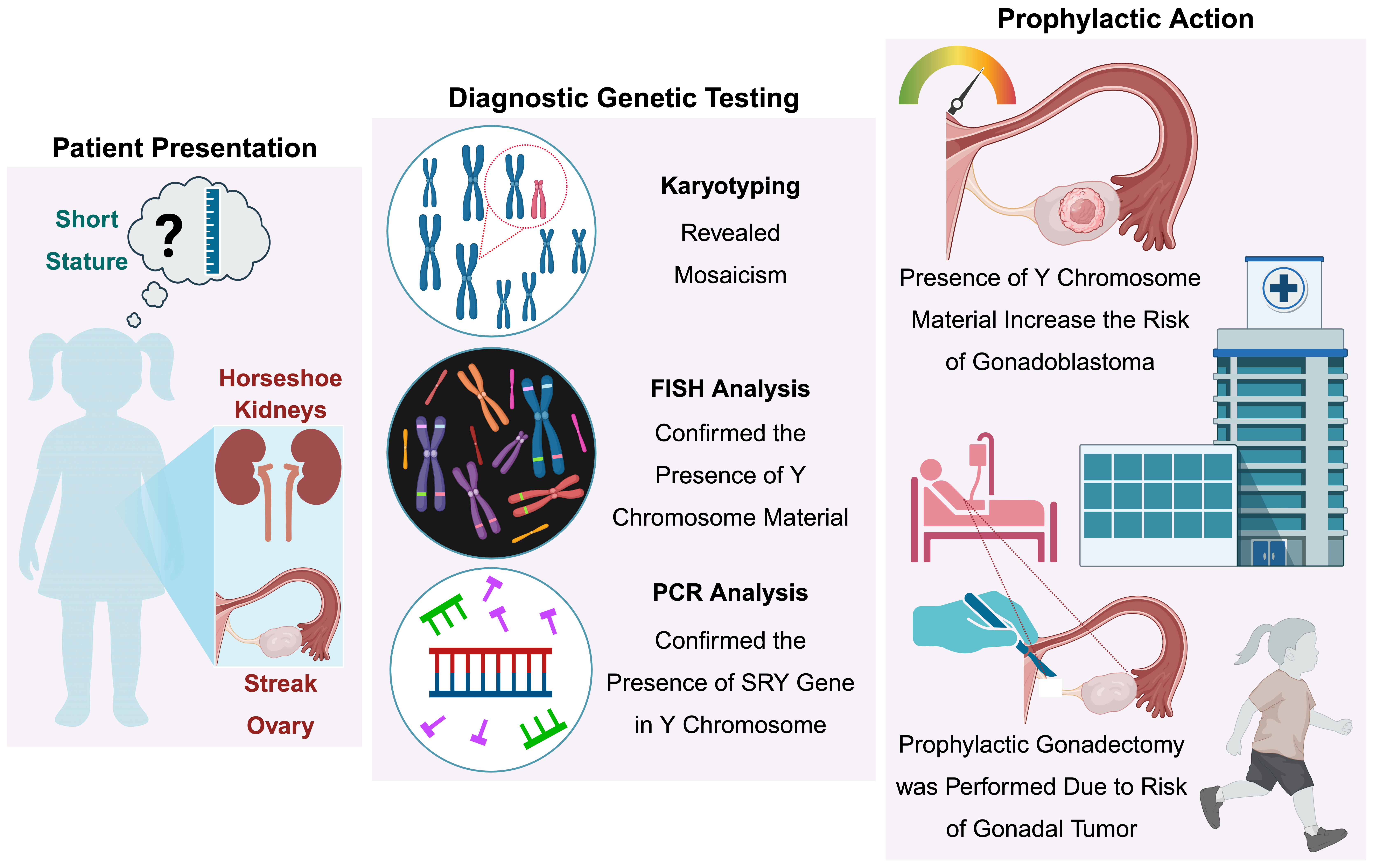
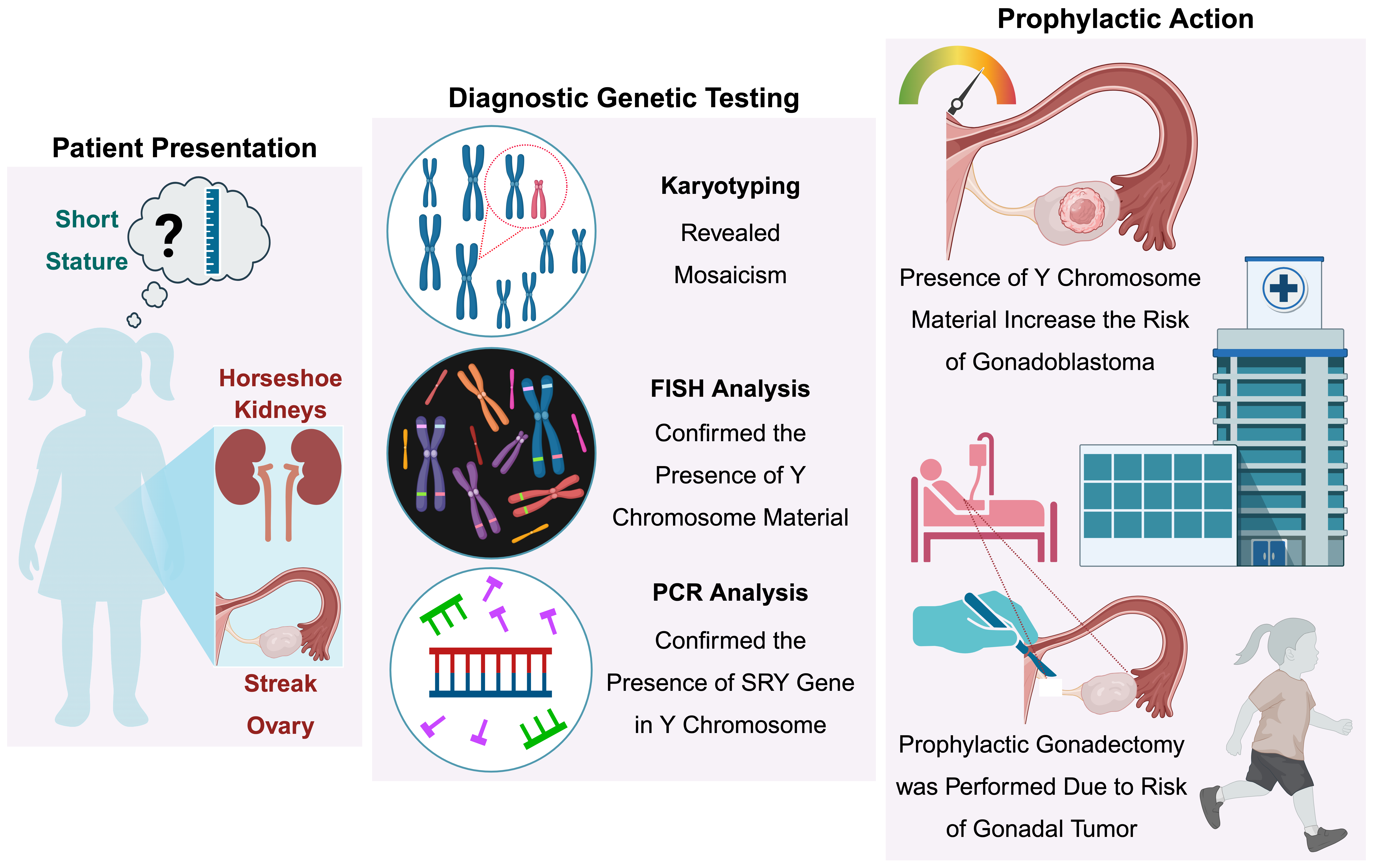
Graphical Abstract
Background
Turner syndrome (TS) is a chromosomal disorder that affects approximately 1 in 2,500 females, with about 50%–60% of TS cases exhibiting monosomy (45,X). The remaining patients demonstrate mosaicism, with one cell line featuring the 45,X karyotype and the other cell lines having structurally abnormal or incomplete X or Y chromosomes1. Structural abnormalities of the X chromosome, such as deletions, inversions, translocations, duplications, ring chromosomes, and isochromosomes, can arise from chromosomal breakage and reunion2. TS is characterized by short stature and gonadal dysgenesis (i.e., impaired development and function of the ovaries). Short stature associated with TS may result from genetic influences or constitutional delays in growth and pubertal development. A key factor contributing to short stature in TS is haploinsufficiency of the short stature homeobox (SHOX) gene, located at the distal end of pseudoautosomal region 1 (PAR1) on the sex chromosomes3,4. More broadly, across the general pediatric population, short stature can arise from a range of etiologies distinct from TS: skeletal dysplasia accounts for approximately 1% of cases, chronic disease for approximately 10%, chromosomal anomalies for approximately 5%, and growth hormone deficiency or receptor insensitivity for 1%–2% of cases, with psychological factors and environmental deprivation contributing in some cases5. In the specific context of TS, however, short stature is attributable to the well-defined mechanism of SHOX haploinsufficiency described above. Furthermore, mosaic patients who additionally carry Y-chromosome material face a distinct clinical concern: an elevated risk of gonadoblastoma.
Patients with mosaic TS, particularly those carrying Y-chromosome material, have a 7%–10% risk of developing gonadoblastoma6. Consequently, careful monitoring is critical for individuals with a 45,X/46,XY karyotype1,7,8. This case study focuses on a 7-year-old girl who presented with unexplained short stature. Cytogenetic analysis and fluorescence in situ hybridization (FISH) testing of peripheral blood lymphocytes revealed an atypical karyotype: 45,X[46]/45,X,der(15)t(Y;15)(p11.32;q10)[4]. Y-autosome translocations are rare chromosomal rearrangements, with an estimated incidence of approximately 1 in 2,000 live births9. In the present case report, we describe the cytogenetic and molecular characterization of this rare Y;15 translocation, confirming its clinical relevance. Based on the observed correlation between karyotype and phenotype, the patient’s family was counseled on appropriate clinical management strategies and associated recurrence risks.
Case Presentation
Patient and Clinical Report
A 7-year-old girl presented to the Kanchi Kamakoti CHILDS Trust Hospital, Chennai, with a chief complaint of unexplained short stature. Chromosomal aberrations were investigated using molecular genetic testing. She was the first child of a healthy, non-consanguineous couple and was born following an uncomplicated pregnancy and vaginal delivery. At the time of her birth, her mother was 19 years old, and her father was 29 years old. Her birth weight was 3.8 kg. At age 7, physical examination revealed that her height and weight were below the third percentile for her age group. Despite her short stature, she exhibited normal intelligence. Physical examination revealed symmetrical features, including joint laxity, mild clinodactyly of the fifth finger, and hypopigmented streaks on her back. Further evaluation, including abdominal ultrasonography, revealed horseshoe-shaped kidneys and a normal uterus, although the ovaries were not visualized. At 8 years of age, diagnostic laparoscopy was performed, revealing bilateral streak ovaries, a hypoplastic uterus, and bilateral hernias. Magnetic resonance imaging (MRI) of the head and thyroid function test results were normal. Additionally, the growth hormone response to clonidine stimulation testing was within the normal range.
Genetic Testing and Counseling
Cytogenetic Analysis
Standard karyotyping was performed at the Department of Medical Genetics at KKCTH after obtaining informed consent from the parents as part of routine diagnostic procedures.
Conventional Cytogenetic Techniques
Human peripheral blood cultures were used to prepare metaphase slides, followed by G-banding using trypsin and Giemsa (GTG-banding) to detect numerical and structural chromosomal abnormalities. Leukocyte culture was initiated by adding 0.5 mL of blood from the proband, collected in a sodium heparin tube (BD Vacutainer, USA), into a 30-mL culture vial containing 5 mL of RPMI 1640 medium, 2 mL of fetal bovine serum (FBS), and 0.2 mL of phytohemagglutinin (PHA). The culture was incubated at 37 °C, and after 69 h, 0.1 mL of colchicine was added. The vials were re-incubated at 37 °C for 12 min to arrest the cells in metaphase. The cell suspension was centrifuged at 1,000 rpm for 10 min to allow the cells to settle. Following removal of the supernatant, 10 mL of pre-warmed, 0.075 M hypotonic potassium chloride (KCl) solution was added, and the vials were incubated at 37 °C for 15 min. The cell suspension was repeatedly centrifuged, and 0.5 mL of fresh Carnoy’s fixative (methanol and glacial acetic acid, 3:1 ratio) was added with gentle mixing until a clear white cell pellet was obtained. Ice-cold cleaned glass slides were used to prepare metaphase spreads. The prepared slides were baked at 58 °C for 24 h.
GTG Banding
Metaphase slides were immersed in 0.005% trypsin for 2 s, followed by treatment with Sørensen's buffer for 5 s. The slides were then stained with Giemsa stain for 5–8 min, rinsed with distilled water, and examined under a light microscope at 100× magnification (BH-2, Olympus, Japan). Metaphase spreads were captured using Cytovision software (version 3.1; Applied Imaging, USA). Cytogenetic analysis of 50 metaphase spreads with a standard band resolution of 450–550 revealed a mosaic karyotype of 45,X[46]/45,X,der(15)t(Y;15)(p11.32;q10)[4], suggesting a TS variant. Among the 50 cells analyzed, 92% (46 cells) exhibited a 45,X karyotype, whereas 8% (4 cells) showed a derivative chromosome 15 resulting from an unbalanced translocation between the Y and 15 chromosomes, consistent with the 45-chromosome count and the absence of a reciprocal derivative Y chromosome (Figure 1A and B). The results were interpreted in accordance with the guidelines of the International System for Human Cytogenomic Nomenclature (ISCN) 2024.
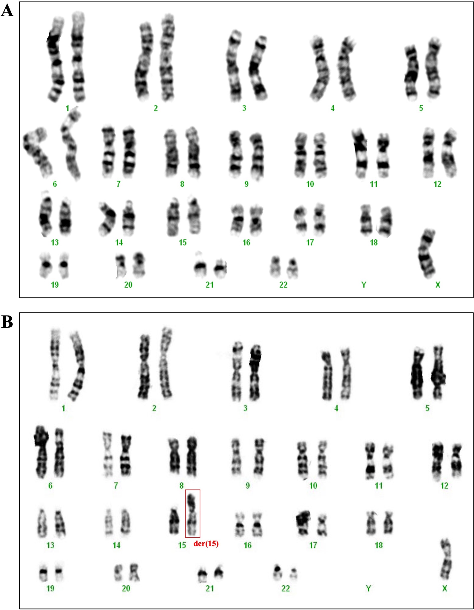
Representative G-banded karyotypes from the patient showing mosaicism: (A) 45,X and (B) 45,X,der(15)t(Y;15)(p11.32;q10).
Molecular Cytogenetic Analysis
Fluorescence in situ hybridization (FISH) was performed on interphase nuclei and metaphase spreads using a three-color CEP X/Y/18 probe (AneuVysion; Cat. No. 05J38-010; Abbott Molecular Vysis, Inc.). This probe targets the centromeric regions of the X (green), Y (red), and 18 (aqua) chromosomes, with chromosome 18 serving as an internal control. All procedures were performed according to the manufacturer's guidelines. An Olympus BX-60 fluorescence microscope and Cytovision software (version 3.1) were used to examine 100 interphase nuclei and 25 metaphase spreads for fluorescence signals. The results showed monosomy X in 88% of the cells and the presence of Y-chromosome material in 12% of the cells (Figure 2). The chromosome 18 signal served as a hybridization efficiency control and was consistently detected in all analyzed interphase nuclei and metaphase spreads. The slightly higher mosaic proportion detected by FISH (12%) than by conventional karyotyping (8%) is expected, as FISH interrogates a substantially larger number of cells (100 interphase nuclei and 25 metaphase spreads) than the 50 metaphase spreads scored by GTG-banding, providing greater sensitivity for detecting low-level mosaicism. Therefore, the two techniques are complementary rather than discordant, with FISH refining and corroborating the cytogenetic findings. In addition, normal male (46,XY) and normal female (46,XX) control slides were processed and hybridized in parallel with the patient's slides to validate the performance, signal specificity, and hybridization efficiency of the X- and Y-chromosome probes.
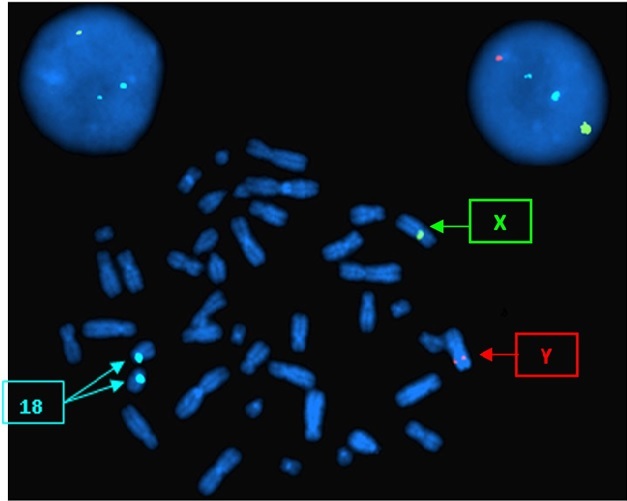
Fluorescence in situ hybridization (FISH) analysis of metaphase spreads and interphase nuclei showing sex chromosome mosaicism. Green signals indicate the X chromosome, red signals indicate the Y chromosome, and aqua signals indicate chromosome 18 (internal control). Monosomy X (45,X) is observed in cells with a single green signal and two aqua signals, while the Y-bearing cell line is identified by the presence of both green (X) and red (Y) signals.
SRY Gene Testing
Polymerase chain reaction (PCR) was performed to detect the sex-determining region Y (SRY) gene. Genomic DNA was isolated from peripheral blood samples using the QIAamp DNA Blood Mini Kit (Qiagen, Germany; Cat. No. 51104) according to the manufacturer’s instructions. Briefly, blood samples were lysed with buffer AL and proteinase K, ethanol was added, and the lysate was applied to a QIAamp spin column. The bound DNA was washed with buffers AW1 and AW2 to remove contaminants, eluted with buffer AE, and stored at −20 °C. DNA concentration and purity were assessed spectrophotometrically, and its integrity was verified by agarose gel electrophoresis.
PCR for the SRY gene was performed using sequence-specific primers (forward: 5′-GAATATTCCCGCTCTCCGCA-3′; reverse: 5′-GCTGGTGCTCCATTCTTGAG-3′). Primer specificity and the expected amplicon size were confirmed in silico using BLAST, which predicted a 470-bp product from the SRY target region. This primer set had been previously validated for sex determination. PCR was performed in a final volume of 20 µL containing 100 ng of genomic DNA, 1× PCR buffer (GeNei Laboratories Pvt. Ltd., India; Cat. No. 0601600051730), 2 mM MgCl₂ (GeNei Laboratories Pvt. Ltd.; Cat. No. 0605710021730), 200 µM dNTPs (GeNei Laboratories Pvt. Ltd.; Cat. No. 652300031730), 0.2–0.5 µM of each primer, and 1 U of Taq DNA polymerase (GeNei Laboratories Pvt. Ltd.; Cat. No. 0601600051730). Amplification was performed in a thermal cycler with an initial denaturation at 94 °C for 5 min, followed by 29 cycles of denaturation at 94 °C for 30 s, annealing at 59 °C for 1 min, and extension at 65 °C for 1 min, with a final hold at 10 °C. DNA samples from healthy males and females were included as positive and negative controls, respectively, to confirm assay specificity. PCR amplicons were resolved on a 2% agarose gel alongside a 100-bp DNA ladder. The ethidium bromide-stained gels were then visualized and imaged. PCR showed positive amplification of the SRY gene (Figure 3), supporting the FISH results and highlighting the importance of molecular confirmation in cases of mosaic TS.
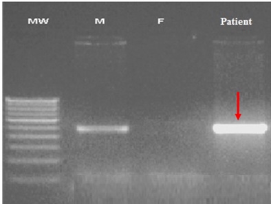
PCR amplification of the SRY gene resolved by agarose gel electrophoresis. The patient sample (indicated by the red arrow) shows a specific 470-bp amplicon, matching the positive control. MW, 100-bp molecular weight marker; M, healthy male genomic DNA (positive control); F, healthy female genomic DNA (negative control).
Genetic Counseling
Based on the observed correlation between the patient's karyotype and phenotype, genetic counseling was provided to the family regarding appropriate clinical management strategies and associated recurrence risks. The presence of a Y chromosome or Y-chromosome fragments in combination with a 45,X karyotype significantly increases the risk of gonadoblastoma, with an estimated likelihood of 7%–10%. Given this elevated risk, the patient’s family was advised to consider prophylactic gonadectomy as a preventive measure.
Discussion and Conclusions
This case report describes a rare chromosomal abnormality in a female child involving a translocation between the Y and 15 chromosomes, occurring in combination with monosomy X. The patient exhibited streak ovaries and underwent prophylactic gonadectomy. Histopathological examination of the excised gonadal tissue revealed no evidence of gonadoblastoma, although the SRY gene was identified on the translocated Y chromosome material. Similar Y-autosome translocations have been documented in multiple cases (Table 1), with an estimated frequency of 1 in 2,000 individuals in the general population9. The likely mechanism of translocation between the Y chromosome and autosomes may result from similarities in the heterochromatic regions of chromosomes 15, 22, and Y10. However, this heterochromatin homology model does not appear to explain the present case. Most previously reported Y;15 translocations (Table 1, cases 1–7) involve breakpoints in the heterochromatic Yq12 region and the heterochromatic short arm of chromosome 15 (15p11, 15p12, or 15p13), consistent with pairing between repetitive heterochromatic sequences. In our patient, however, the breakpoints mapped to Yp11.32 (a euchromatic region on the short arm of the Y chromosome) and to 15q10 (the centromeric region of chromosome 15). This euchromatic/centromeric breakpoint configuration is atypical for the heterochromatin-mediated mechanism classically invoked for Y-autosome translocations and suggests that the rearrangement in this case may have arisen through a different mechanism, such as non-allelic homologous recombination between euchromatic repeat elements or non-homologous end joining/microhomology-mediated double-strand break repair. This distinction highlights that Y;15 translocations may arise through more than one mechanistic pathway, depending on the specific breakpoint regions involved.
Previously reported translocations between the Y chromosome and autosomes. Note: All karyotype designations are presented using their original nomenclature to maintain historical consistency.
| No. | Karyotype | Specimen Type | Phenotype | References | |
|---|---|---|---|---|---|
| 1 | 46,XY,der(15),t(Y;15)(q12;p13) | Not performed | Umbilical cord blood, lymphocytes | Normally developed | |
| 2 | 46,XX,der(15),t(Y;15)(q12;p13) | Not performed | Umbilical cord blood, lymphocytes | Normally developed | |
| 3 | 46,XX,der(15),t(Y;15)(q12;p11) | Not performed | Peripheral leukocytes, cultured skin fibroblasts, EBV-transformed lymphoblasts | Familial translocation, phenotypically normal | |
| 4 | 46,XX,der(15),t(Y;15)(q12;p11) | Not performed | PHA-stimulated whole blood cultures | Normally developed | |
| 5 | 46,X,t(Y;15)(q12;p13)/45,X | Positive | Blood lymphocytes, skin | Late onset of menarche | |
| 6 | 46,XX,der(15)t(Y;15)(q12;p13).ish der(15)t(Y;15)(q12;p13)(DYZ1,DYZ3y,D15Z1) | Not performed | Blood | Two pregnancy losses with trisomy 15 and one with tetraploidy | |
| 7 | 46,XX,der(15)t(Y;15)(q12;p12)pat. ish der(15)(DYZ1+1) | Positive | Uncultured amniotic fluid cells | Normally developed |
The presence of a Y chromosome or a Y-chromosome fragment in a patient with a 45,X karyotype significantly increases the risk of gonadoblastoma, estimated at 7%–10%6. This can lead to virilization of the female genitalia, which may present as ambiguous genitalia16. Based on this information, the family was advised to consider preventive gonadectomy. Individuals with a translocation between the Y chromosome and an autosome can present with a range of distinct clinical phenotypes17. For instance, Chen et al.17 described a phenotypically normal adult male carrier of a unique Y;14 translocation, identified incidentally during an infertility work-up, in whom the rearrangement was inherited and associated with azoospermia rather than a TS phenotype. This contrasts with our patient, in whom the Y;15 translocation occurred in the context of 45,X mosaicism, was apparently de novo, and manifested as short stature and gonadal dysgenesis rather than infertility in a phenotypically male carrier. Taken together, these two cases illustrate that the clinical consequence of a Y-autosome translocation depends heavily on the sex chromosome constitution and cell-line distribution in which it occurs, rather than on the identity of the autosomal partner chromosome alone. Despite the absence of a second X chromosome, the patient did not exhibit typical somatic features of TS, with the only observed characteristics being unexplained short stature and streak ovaries. Karyotyping revealed a Turner mosaic variant, which accounted for her short stature. Subsequent molecular investigations, including FISH and SRY gene analysis, enabled effective counseling regarding clinical care decisions and associated recurrence concerns. The present case emphasizes the importance of integrating FISH, molecular analysis, and conventional G-banded cytogenetic approaches for the diagnosis of unexplained short stature associated with mosaic Turner variants.
Y-autosome translocations are frequently correlated with male infertility and azoospermia, whereas the incidence of Y-autosome translocations in females is extremely rare. This case report presents a chromosomal aberration of 45,X[46]/45,X,der(15)t(Y;15)(p11.32;q10)[4] in a female child with unexplained short stature but without other characteristic physical signs of TS. Although clinically significant, this case report has some limitations. These include our inability to employ additional techniques, such as C-banding or array comparative genomic hybridization (aCGH), for a more comprehensive understanding of the chromosomal alterations in the derivative chromosome 15. Additionally, we were unable to perform parental testing to ascertain the origin of this chromosomal abnormality due to the parents' unwillingness to undergo chromosomal analysis. A de novo event is associated with a negligible risk of recurrence in subsequent parental pregnancies, except in rare cases of parental germline mosaicism17. In contrast, the identification of a maternal or paternal carrier state confers a substantial and predictable risk of transmission to offspring, increases the likelihood of unbalanced gametes, and may result in recurrent pregnancy loss or reproductive failure in future gestations5. The present case strongly demonstrates that combining conventional cytogenetics with FISH and SRY gene testing is essential for the detection of Y-chromosome material, thereby enabling life-saving preventive measures such as prophylactic gonadectomy.
Declarations
Abbreviations
aCGH: array comparative genomic hybridization; BLAST: Basic Local Alignment Search Tool; bp: base pair; DNA: deoxyribonucleic acid; dNTPs: deoxynucleotide triphosphates; EBV: Epstein-Barr virus; FBS: fetal bovine serum; FISH: fluorescence in situ hybridization; GTG-banding: G-banding using trypsin and Giemsa; ISCN: International System for Human Cytogenomic Nomenclature; KCl: potassium chloride; KKCTH: Kanchi Kamakoti CHILDS Trust Hospital; MgCl₂: magnesium chloride; MRI: magnetic resonance imaging; PCR: polymerase chain reaction; PHA: phytohemagglutinin; RPMI: Roswell Park Memorial Institute; SRY: sex-determining region Y gene; SHOX: short stature homeobox gene; PAR1: pseudoautosomal region 1; TS: Turner syndrome
Acknowledgments
We thank the study participants for providing essential samples and adhering to ethical standards. We sincerely thank the heads and staff of the research and laboratory facilities at several institutions, including the Medical Genetics Department at KKCTH in Chennai and the Human Genetics Department at SRIHER, Chennai. Additionally, we thank all the reagent and equipment suppliers for their unwavering support and technical expertise, which enabled us to obtain accurate and meaningful results. Graphical abstract created in BioRender. SHRESTHA, R. (2026) .
Author’s contributions
MJK and KG conceptualized this study. MJK conducted the investigation, analyzed the data, and drafted the manuscript. MJK, KG, TK, LR, RS, RSh, AK, PD, and SD contributed to the formal analysis, validation, and editing of the manuscript. All authors have read and approved the final version of the manuscript.
Funding
This study was self-funded and did not receive any external financial support.
Availability of data and materials
All data generated or analyzed during this study are included in this published article.
Ethics approval and consent to participate
This study was approved by the Ethics Committee of the SRM Medical College Hospital and Research Centre (Ethics Clearance number: 1281/IEC/2017). Written informed consent was obtained from the patient’s parents/guardians for participation in this study.
Consent for publication
Written informed consent was obtained from the patient’s parents/guardians for the publication of this case report and any accompanying images.
Declaration of generative AI and AI-assisted technologies in the writing process
The authors declare that no generative AI or AI-assisted technologies were used in the writing or editing of this manuscript.
Competing interests
The authors declare that they have no competing interests.