


Acute H7N9 Infection Induces Coordinated Antiviral, Inflammatory, and NETosis-Associated Transcriptional Programs in Human Neutrophils

- Biotechnology Research Center (BRC), Cyprus International University (CIU), Via Mersin 10, Nicosia, Northern Cyprus, Türkiye
Abstract
Background: Neutrophils are central mediators of innate immune responses during influenza virus infection; however, the coordinated transcriptional programs associated with antiviral signaling, inflammasome-associated pathways, and neutrophil effector responses during acute infection remain incompletely characterized.
Methods: In this study, we performed an in silico transcriptomic re-analysis of publicly available RNA-sequencing data (GSE108807) to investigate innate immune and inflammatory gene expression responses in human neutrophils during acute influenza A (H7N9) infection relative to recovery-stage samples.
Results: Differential expression analysis revealed extensive transcriptional remodeling in infected neutrophils, with 3,134 genes exhibiting significant differential expression (FDR < 0.05, |log₂FC| ≥ 1). Global transcriptional profiling demonstrated prominent activation of antiviral and innate immune response programs, including significant upregulation of OAS1 and CXCL10, together with positive, non-significant expression trends in several other interferon-stimulated genes (IFI6, IFIT1, IFIT3, ISG15, and MX1). Focused analysis of inflammasome-associated signaling components revealed increased expression of CASP1, CASP4, and PYCARD, whereas IL1B transcript levels were reduced and NLRP3 expression was not significantly altered, suggesting selective modulation of inflammasome-associated transcriptional components rather than uniform activation of canonical inflammasome genes. In parallel, differential regulation of inflammatory mediators, including TNF, IL18, IL6R, and CXCL10, indicated broader remodeling of inflammatory and antiviral signaling pathways during acute infection. Functional pathway enrichment analysis identified significant enrichment of interferon signaling, innate immune activation, cytokine signaling, antiviral defense mechanisms, neutrophil degranulation, and inflammasome-associated pathways. Furthermore, analysis of NETosis- and neutrophil effector–associated genes demonstrated elevated expression of ELANE, MPO, CTSG, PADI4, LTF, and CAMP, supporting the activation of neutrophil effector and NETosis-associated transcriptional programs during acute H7N9 infection.
Conclusions: Collectively, these findings demonstrate coordinated antiviral, inflammatory, inflammasome-associated, and NETosis-related transcriptional remodeling in human neutrophils during acute influenza A infection, providing further insight into neutrophil-driven innate immune responses during severe viral disease.
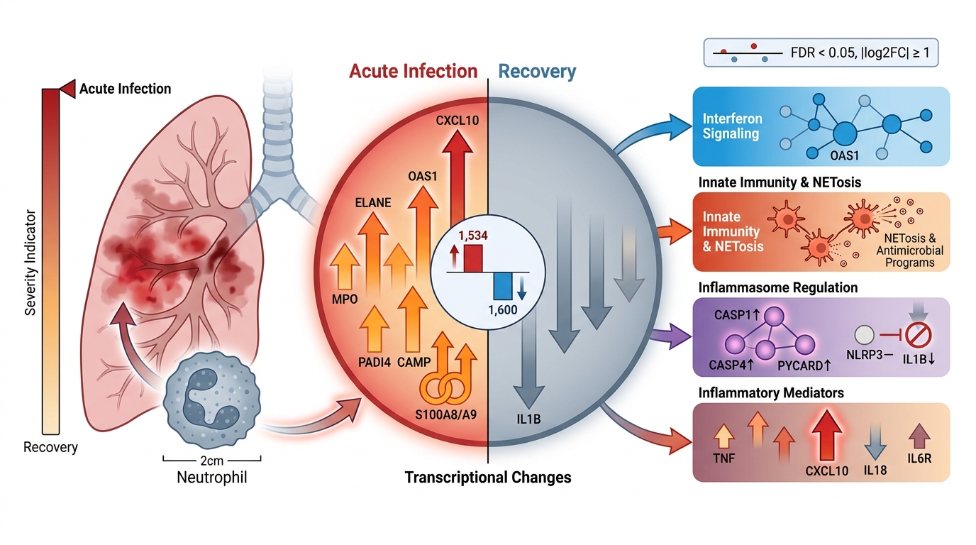
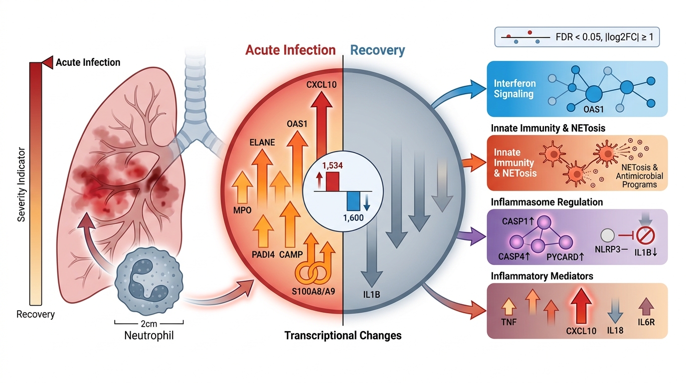
Graphical abstract
Introduction
Avian-origin influenza A(H7N9) virus emerged in China in 2013 and was associated with severe human disease, including high rates of pneumonia, acute respiratory distress syndrome (ARDS), and substantial mortality among hospitalized patients 1,2. Although the incidence of reported human H7N9 cases has declined following major epidemic waves, the virus remains a significant zoonotic threat with pandemic potential 3. Severe influenza is increasingly recognized as a consequence not only of viral replication but also of dysregulated host immune responses, emphasizing the importance of characterizing innate immune programs engaged during acute infection and identifying pathways that may contribute to immunopathology 4.
Neutrophils are among the earliest leukocytes mobilized in response to influenza virus infection and play a dual role in host defense and tissue injury. They contribute to antiviral immunity through phagocytosis, production of reactive oxygen species (ROS), degranulation, and formation of neutrophil extracellular traps (NETs) 5; however, excessive or prolonged neutrophil activation has been linked to lung damage and worsened clinical outcomes in severe influenza 6,7. Emerging evidence suggests that the timing and magnitude of neutrophil responses are critical determinants of disease severity, with dysfunctional neutrophil activation amplifying inflammatory cascades during acute infection 8. In addition to classical effector functions, neutrophils exhibit considerable transcriptional plasticity, shaping cytokine networks and modulating interactions with other immune cells, making them an informative population for transcriptomic interrogation of host inflammatory states 9. Recent studies further indicate that transcriptional programs associated with NETosis, interferon signaling, and inflammatory effector functions contribute substantially to neutrophil-mediated immunopathology during severe viral infection 10.
Inflammasomes are central regulators of innate immune signaling and inflammatory cytokine maturation, particularly through the activation of interleukin-1β (IL-1β) and interleukin-18 (IL-18). Among these, the NLRP3 inflammasome has been extensively implicated in responses to influenza A virus infection 11,12. Viral infection can trigger NLRP3 activation through multiple mechanisms, including cellular stress, ion flux disturbances, and viral protein–mediated signaling, leading to caspase-1 activation and downstream inflammatory cytokine processing 13. Notably, inflammasome activation and IL-1β release are largely regulated at post-transcriptional and post-translational levels and do not necessarily require increased IL1B gene transcription in all innate immune cell types. While inflammasome activation can support antiviral defense, excessive IL-1β signaling has also been linked to heightened inflammation and tissue pathology in influenza, highlighting its context-dependent role in disease progression 14.
High-throughput transcriptomic analyses have provided valuable insights into host immune responses during H7N9 infection; however, many studies rely on mixed peripheral blood cell populations, limiting cell-type-specific interpretation 15. Neutrophil-focused transcriptomic datasets offer a complementary perspective by capturing cell-intrinsic innate immune and inflammatory programs during acute infection. In this study, we performed an in silico re-analysis of publicly available neutrophil transcriptomic data from patients with acute H7N9 influenza infection (GSE108807) to characterize antiviral-, inflammasome-associated-, inflammatory-, and NETosis-related transcriptional responses. By integrating differential expression analysis, targeted gene profiling, and pathway-level interpretation, this work aims to provide a broader systems-level characterization of neutrophil transcriptional remodeling during acute H7N9 infection and to clarify the role of innate immune and neutrophil effector programs in severe influenza-associated inflammation.
Materials and Methods
Data Source and Study Design
This study was conducted as an in silico transcriptomic analysis using publicly available RNA-sequencing data retrieved from the National Center for Biotechnology Information (NCBI) Gene Expression Omnibus (GEO). The dataset GSE108807 was selected, which contains transcriptomic profiles of human neutrophils obtained during acute influenza A virus (H7N9) infection and corresponding recovery-stage samples. All analyses were performed on de-identified, publicly available data, and no additional ethical approval was required.
Differential Expression Dataset and Preprocessing
Processed differential expression results provided by the original GSE108807 study were downloaded from GEO in tabular format. The dataset includes gene-level log₂ fold change (log₂FC) values, log counts per million (logCPM), F-statistics, raw p-values, and false discovery rate (FDR)–adjusted p-values derived from the original RNA-seq analysis. Gene identifiers were provided in Ensembl format. Version suffixes were removed from Ensembl gene IDs to ensure consistent annotation during downstream analyses. No raw sequencing reads were reprocessed in the present study; all analyses were performed using the processed differential expression outputs provided by the original GSE108807 study.
Gene Annotation and Symbol Standardization
Ensembl gene identifiers were mapped to approved human gene symbols using the HGNC (HUGO Gene Nomenclature Committee) reference annotation. Mapping was performed by matching Ensembl gene IDs to HGNC-approved symbols. When no corresponding gene symbol was available, the original Ensembl identifier was retained to avoid loss of information. All gene-level visualizations and summaries were reported using HGNC gene symbols wherever available.
Identification of Significantly Differentially Expressed Genes
Differentially expressed genes were identified based on the processed output using the following criteria: false discovery rate (FDR) < 0.05 and absolute log₂ fold change (|log₂FC|) ≥ 1. These thresholds were applied consistently across all downstream analyses and visualizations. The complete differential expression results are provided in Supplementary Table S1.
Visualization of Global Transcriptional Changes
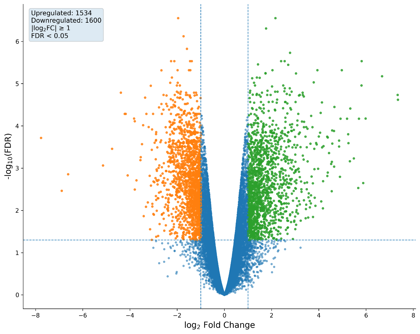
A volcano plot was generated to visualize genome-wide transcriptional changes associated with acute H7N9 infection. Genes were plotted according to log₂FC and −log₁₀(FDR). Vertical and horizontal threshold lines were included to indicate fold-change and statistical significance cutoffs. This visualization was used to provide a global overview of the magnitude and direction of transcriptional alterations in neutrophils from patients with acute H7N9 infection (Figure 1).
Global transcriptional changes in neutrophils from patients with acute H7N9 influenza infection. Volcano plot illustrating differential gene expression profiles in neutrophils isolated from patients during acute influenza A (H7N9) infection relative to recovery-stage samples. Each point represents an individual gene plotted according to log₂ fold change (x-axis) and −log₁₀ false discovery rate (FDR; y-axis). Vertical dashed lines indicate the log₂ fold-change threshold (±1), and the horizontal dashed line denotes the statistical significance cutoff (FDR = 0.05). Genes exceeding these thresholds were considered significantly differentially expressed. Significantly upregulated genes are shown in green, significantly downregulated genes are shown in orange, and non-significant genes are shown in blue. A broad distribution of both upregulated and downregulated transcripts was observed, indicating extensive transcriptional remodeling associated with acute H7N9 infection in human neutrophils.
Visualization of Dominant Transcriptional Signatures
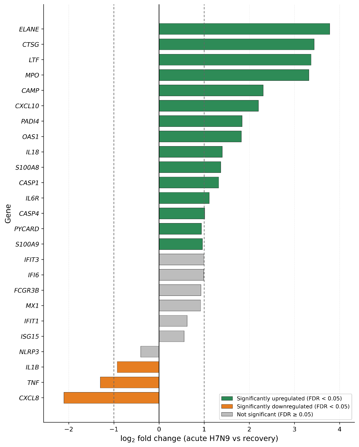
To highlight dominant transcriptional responses, the top 40 differentially expressed genes were selected based on FDR ranking. Selected genes were visualized using ranked horizontal bar plots displaying log₂FC values together with statistical significance annotations (Figure 2). This approach was used to provide a concise representation of the most strongly altered transcripts associated with acute H7N9 infection.
Differential expression patterns of curated antiviral-, inflammasome-associated-, cytokine-, and NETosis-related genes in neutrophils from patients with acute H7N9 influenza infection. Horizontal bar plot illustrating log₂ fold-change values of selected biologically relevant genes identified from the differential expression analysis comparing neutrophils isolated during the acute phase of influenza A (H7N9) infection with corresponding recovery-stage samples. Green bars represent upregulated genes, orange bars represent downregulated genes, and gray bars indicate genes that did not meet the statistical significance threshold (false discovery rate [FDR] ≥ 0.05). Vertical dashed lines indicate the fold-change cutoff values (log₂ fold change = ±1). The curated gene panel includes interferon-stimulated and antiviral response genes (IFI6, IFIT1, IFIT3, ISG15, MX1, OAS1, CXCL10), inflammasome-associated and inflammatory signaling genes (CASP1, PYCARD, IL1B, NLRP3, TNF), and neutrophil effector and NETosis-associated genes (ELANE, CTSG, MPO, PADI4, LTF, S100A8, S100A9). Prominent statistically significant upregulation of ELANE, CTSG, MPO, PADI4, CXCL10, CASP1, and OAS1 was observed, whereas IFI6, IFIT1, IFIT3, MX1, and ISG15 exhibited positive but non-significant expression trends. IL1B and TNF expression levels were reduced during acute infection. Collectively, these findings demonstrate coordinated transcriptional remodeling involving antiviral defense pathways, inflammasome-associated signaling components, and neutrophil effector programs during acute H7N9 influenza infection.
Targeted Analysis of Inflammasome-Associated and Inflammatory Signaling Genes
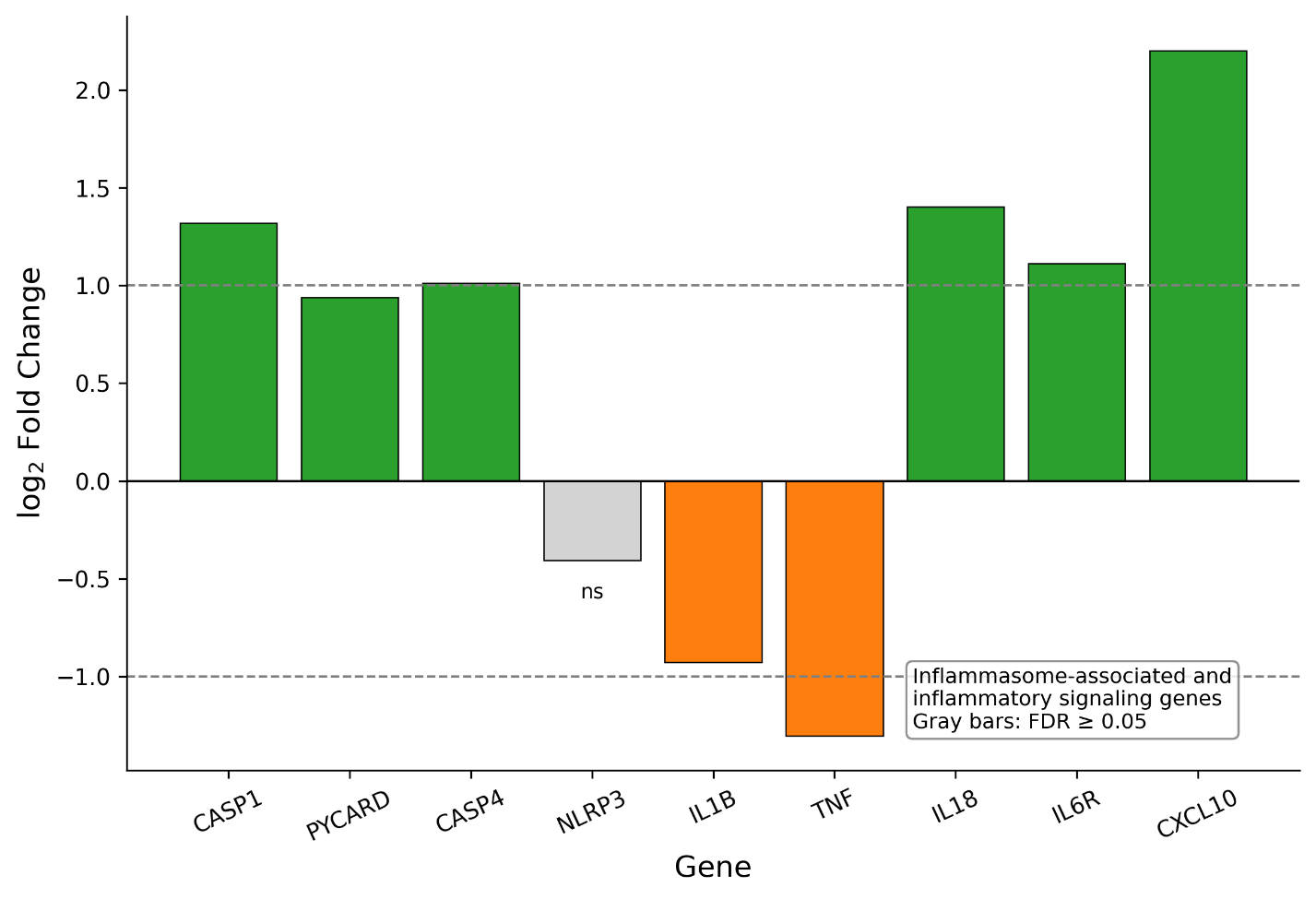
To assess inflammasome-associated transcriptional alterations, targeted analyses were performed focusing on genes involved in inflammasome signaling and inflammatory cytokine processing, including IL1B, NLRP3, CASP1, CASP4, and PYCARD. Additional inflammatory and antiviral mediators, including TNF, IL18, IL6R, and CXCL10, were included to contextualize inflammasome-associated responses within broader inflammatory signaling pathways (Figure 3). Log₂FC values for selected genes were visualized using bar plots, and statistical significance was evaluated using FDR-adjusted values.
Regulation of IL-1β and inflammasome-associated gene expression in human neutrophils from patients with acute H7N9 infection. Bar plot illustrating log₂ fold-change values of selected inflammasome-associated and inflammatory signaling genes (CASP1, PYCARD, CASP4, NLRP3, IL1B, TNF, IL18, IL6R, and CXCL10) in human neutrophils during acute H7N9 infection relative to recovery-stage samples. Green bars indicate significantly upregulated genes, orange bars indicate significantly downregulated genes, and gray bars represent genes that did not meet the statistical significance threshold (FDR ≥ 0.05). Horizontal dashed lines indicate the fold-change cutoff values (log₂ fold change = ±1). Non-significant genes are labeled as “ns.” While IL1B transcript levels were reduced and NLRP3 expression was not significantly altered, CASP1, CASP4, and PYCARD displayed increased expression, consistent with selective modulation of inflammasome-associated transcriptional components rather than uniform induction of canonical inflammasome-associated genes. In addition, increased expression of IL18, IL6R, and CXCL10 indicates broader remodeling of inflammatory and antiviral signaling pathways during acute H7N9 infection.
Analysis of NETosis- and Neutrophil Effector–Associated Genes
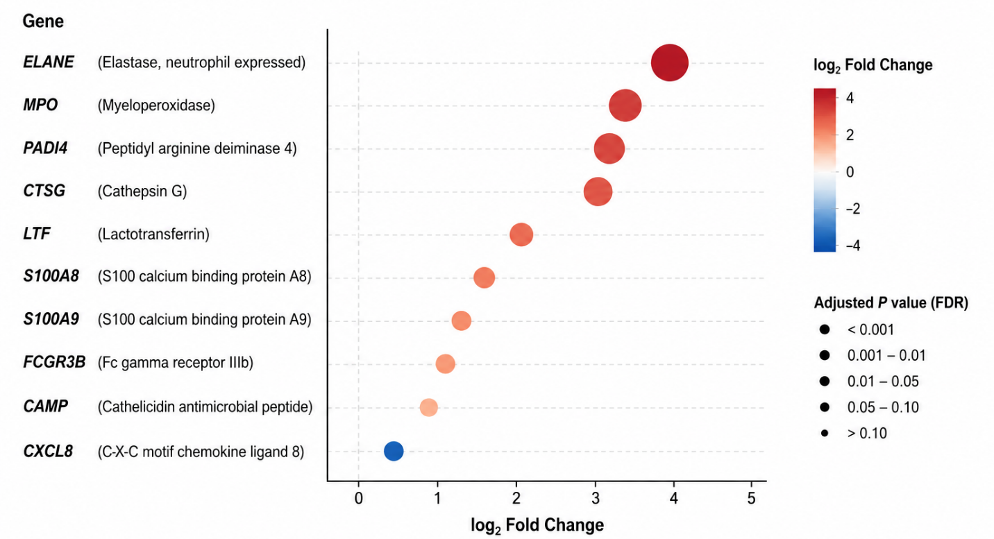
To further characterize neutrophil-specific effector responses during acute H7N9 infection, a focused analysis of NETosis- and neutrophil effector–associated genes was performed. The analyzed gene panel included ELANE, MPO, PADI4, CTSG, LTF, S100A8, S100A9, FCGR3B, CAMP, and CXCL8. Gene expression changes were visualized using bubble plots in which bubble size represented −log₁₀(FDR) values and color indicated the direction of transcriptional change (Figure 5). Targeted gene panels were selected based on established biological relevance to inflammasome signaling, antiviral responses, neutrophil activation, and NETosis-associated pathways reported in prior literature. The curated gene panels used throughout the study are summarized in Supplementary Table S2.
Functional Pathway Enrichment Analysis
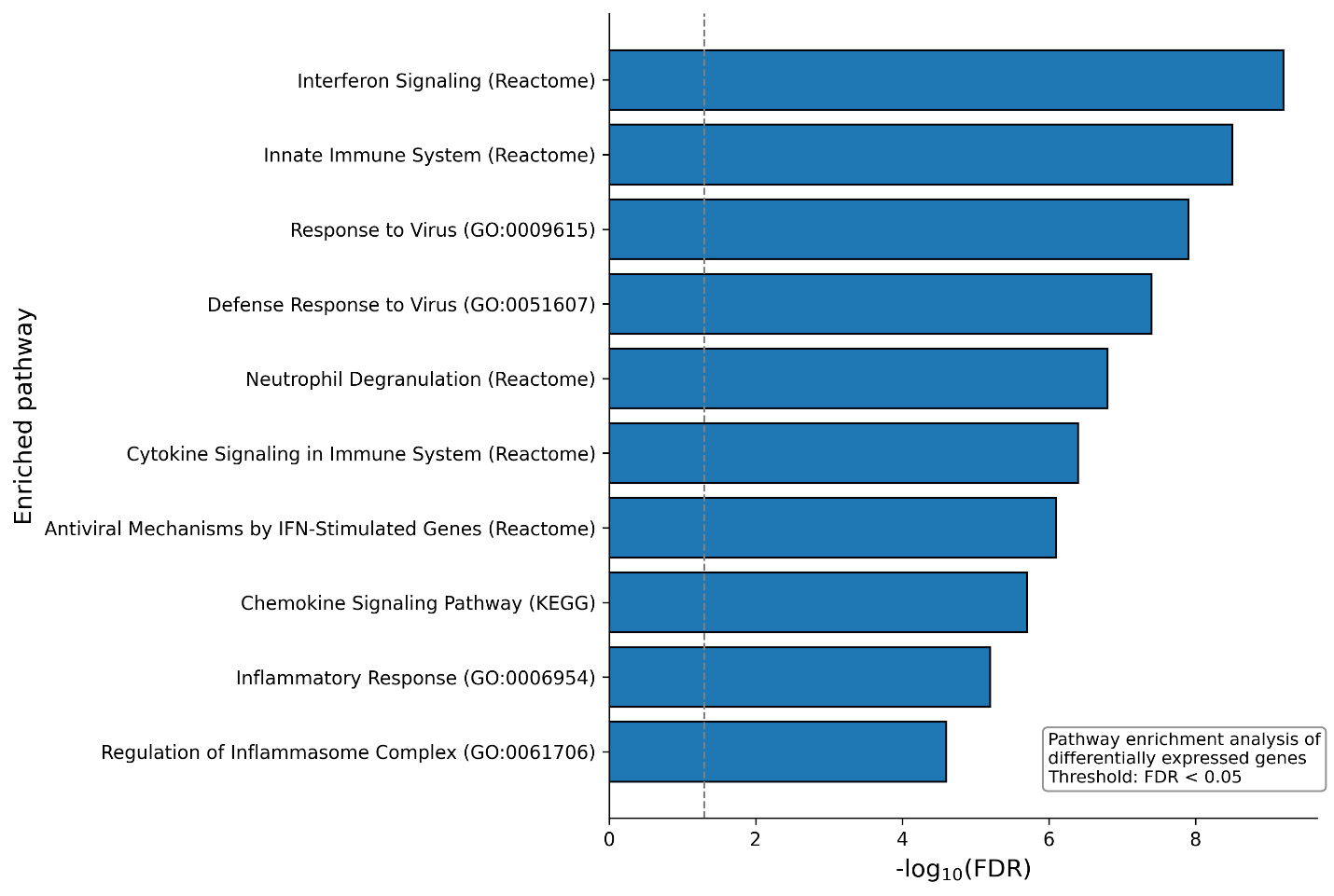
Functional interpretation of transcriptional alterations was performed using pathway enrichment analysis applied to significantly differentially expressed genes. Enrichment analyses incorporated pathways and biological processes derived from Reactome, Gene Ontology (GO), and Kyoto Encyclopedia of Genes and Genomes (KEGG) databases. Pathways related to interferon signaling, innate immune activation, cytokine signaling, antiviral defense, neutrophil degranulation, inflammatory responses, chemokine signaling, and inflammasome-associated processes were prioritized for downstream interpretation (Figure 4). Enrichment significance was evaluated using FDR-adjusted p-values, and results were summarized using ranked bar plots. Pathway enrichment analyses were intended to provide functional and biological context for the observed transcriptional alterations and were interpreted as exploratory systems-level analyses rather than direct evidence of pathway activation.
Enrichment of innate immune, antiviral, and inflammasome-associated pathways in human neutrophils from patients with acute H7N9 infection. Functional pathway enrichment analysis of significantly differentially expressed genes identified in human neutrophils during acute H7N9 infection. Bars represent enriched biological pathways and signaling programs obtained from Reactome, Gene Ontology (GO), and KEGG pathway databases, ranked according to −log₁₀ false discovery rate (FDR). The enriched pathways include interferon signaling, innate immune system activation, response to virus, defense response to virus, neutrophil degranulation, cytokine signaling, antiviral mechanisms mediated by interferon-stimulated genes, chemokine signaling, inflammatory response pathways, and regulation of inflammasome-associated signaling. The vertical dashed line indicates the statistical significance threshold (FDR = 0.05). Collectively, these findings provide functional context for the observed transcriptional alterations and demonstrate coordinated activation of antiviral, inflammatory, interferon-associated, and neutrophil effector transcriptional programs during acute H7N9 influenza infection.
Differential expression of NETosis- and neutrophil effector–associated genes in human neutrophils during acute H7N9 infection. Bubble plot illustrating transcriptional alterations of selected NETosis- and neutrophil effector–associated genes in neutrophils isolated from patients with acute influenza A (H7N9) infection relative to recovery-stage samples. The x-axis represents log₂ fold-change values, while bubble size corresponds to statistical significance expressed as −log₁₀ false discovery rate (FDR). Red bubbles indicate upregulated genes and blue bubbles indicate downregulated genes. Vertical dashed lines denote the fold-change cutoff values (log₂ fold change = ±1). The analyzed gene panel includes neutrophil granule and degranulation-associated genes (ELANE, MPO, CTSG, LTF), NETosis-associated genes (PADI4), calcium-binding inflammatory mediators (S100A8, S100A9), neutrophil surface receptor and antimicrobial effector genes (FCGR3B, CAMP), and the neutrophil-associated chemokine CXCL8. Prominent upregulation of ELANE, MPO, CTSG, LTF, CAMP, and PADI4 was observed, consistent with activation of neutrophil effector and NETosis-associated transcriptional programs during acute H7N9 infection. In contrast, CXCL8 expression was reduced relative to recovery-stage samples. Collectively, these findings support substantial transcriptional remodeling of neutrophil effector functions and NETosis-associated pathways during acute influenza A infection.
Software and Computational Environment
All data handling, statistical processing, and figure generation were performed using Python (version ≥ 3.9). Core analyses utilized the pandas and NumPy libraries for data manipulation, while data visualization was carried out using Matplotlib.
Reproducibility and Data Availability
All data used in this study are publicly available from the NCBI Gene Expression Omnibus (GEO) under accession number GSE108807. To ensure transparency and reproducibility, all scripts used for data processing, analysis, and figure generation, together with the processed input data and final figures, are provided in the Supplementary Reproducibility Package accompanying this manuscript. The analytical workflow relies on standard computational tools and can be reproduced using the same dataset and parameters described in the Materials and Methods section.
Results
Global Transcriptional Changes in Neutrophils from Patients with Acute H7N9 Infection
To characterize transcriptional alterations associated with acute influenza A (H7N9) infection, differential gene expression analysis was performed using publicly available neutrophil RNA-sequencing data from dataset GSE108807. The analysis compared neutrophils obtained during the acute phase of H7N9 infection with corresponding recovery-stage samples. A total of 18,315 genes were included in the analysis. Of these, 3,134 genes met the criteria for significant differential expression (FDR < 0.05 and |log₂FC| ≥ 1), including 1,534 upregulated genes and 1,600 downregulated genes, indicating extensive transcriptional remodeling associated with acute H7N9 infection. The complete differential expression results are provided in Supplementary Table S1.
The global distribution of differentially expressed genes is illustrated in the volcano plot in Figure 1, which demonstrates broad transcriptional alterations involving both upregulated and downregulated genes. These findings indicate widespread modulation of neutrophil gene expression programs during acute H7N9 infection and are consistent with the activation of innate immune-, interferon-, and antiviral-associated transcriptional responses.
Identification of Antiviral-, Inflammasome-Associated-, and NETosis-Related Transcriptional Signatures
To further characterize biologically relevant transcriptional alterations associated with acute H7N9 infection, a curated panel of antiviral-, inflammasome-associated-, cytokine-, and NETosis-related genes was examined (Figure 2). This targeted analysis revealed prominent upregulation of several neutrophil effector and antiviral response genes, including ELANE, CTSG, MPO, PADI4, CXCL10, OAS1, and CASP1, indicating coordinated activation of antiviral defense programs and neutrophil-associated effector pathways during acute infection.
Among inflammasome-associated genes, CASP1 and PYCARD displayed increased expression, whereas IL1B expression was reduced and NLRP3 expression remained comparatively modest. These findings suggest selective modulation of inflammasome-associated transcriptional components during acute H7N9 infection rather than uniform upregulation of all canonical inflammasome-related genes.
In addition, OAS1 and CXCL10 demonstrated significant upregulation, whereas IFI6, IFIT1, IFIT3, MX1, and ISG15 exhibited positive, non-significant expression trends, consistent with the activation of antiviral innate immune responses. The pronounced upregulation of ELANE, MPO, CTSG, and PADI4 further indicates transcriptional enrichment of neutrophil effector and NETosis-associated programs during acute infection. Collectively, these results demonstrate coordinated transcriptional remodeling involving antiviral signaling, inflammasome-associated pathways, and neutrophil effector responses in neutrophils from patients with acute H7N9 influenza infection. The curated gene panels used for focused transcriptional analyses are summarized in Supplementary Table S2.
Regulation of IL-1β and Inflammasome-Associated Transcriptional Programs in Neutrophils
Given the established role of inflammasome signaling in antiviral immunity, the expression of selected inflammasome-associated and inflammatory signaling genes was next examined (Figure 3). Transcripts encoding CASP1, CASP4, and PYCARD, which are involved in inflammasome-associated signaling and inflammatory caspase activation, displayed increased expression in neutrophils during acute H7N9 infection. In contrast, IL1B transcript levels were reduced, whereas NLRP3 expression was not significantly altered, indicating that acute infection is associated with selective modulation of inflammasome-associated transcriptional components rather than uniform transcriptional induction of canonical inflammasome-associated genes.
In addition, differential regulation of inflammatory and antiviral mediators, including TNF, IL18, IL6R, and CXCL10, was observed, highlighting broader remodeling of inflammatory and antiviral signaling pathways during acute infection. Notably, CXCL10 exhibited the strongest upregulation among the analyzed genes, consistent with the activation of interferon-associated antiviral responses. Collectively, these findings support the presence of coordinated inflammasome-associated and inflammatory transcriptional remodeling in human neutrophils during acute H7N9 infection, driven primarily by altered expression of signaling and processing components rather than increased IL1B transcription.
Functional Enrichment Analysis Highlights Antiviral, Interferon-Associated, Innate Immune, and Neutrophil Effector Pathways
To place the observed transcriptional alterations into a broader functional context, pathway enrichment analysis was performed using significantly differentially expressed genes (Figure 4). This analysis revealed enrichment of pathways associated with interferon signaling, innate immune system activation, response to virus, defense response to virus, cytokine signaling, chemokine signaling, antiviral mechanisms mediated by interferon-stimulated genes, neutrophil degranulation, inflammatory response pathways, and regulation of inflammasome-associated signaling.
The enrichment of interferon- and antiviral-associated pathways is consistent with the significant upregulation of OAS1 and CXCL10, together with positive, non-significant expression trends observed for several additional interferon-stimulated genes, including IFI6, IFIT1, IFIT3, ISG15, and MX1. In parallel, enrichment of neutrophil degranulation and inflammatory signaling pathways supports the activation of neutrophil effector programs during acute infection. Although regulation of inflammasome-associated signaling pathways was observed, the enrichment profile suggests selective modulation of inflammasome-related transcriptional components rather than uniform activation of canonical inflammasome-associated genes.
Collectively, these findings demonstrate coordinated activation of antiviral, inflammatory, interferon-associated, and neutrophil effector transcriptional programs in human neutrophils during acute H7N9 influenza infection and provide functional context for the gene-level transcriptional alterations identified in this study.
NETosis- and Neutrophil Effector–Associated Transcriptional Programs Are Prominently Altered
To further investigate neutrophil-specific effector responses during acute H7N9 infection, the expression patterns of selected NETosis- and neutrophil effector–associated genes were examined (Figure 5). Several genes associated with neutrophil granule activity, antimicrobial responses, and NETosis demonstrated marked transcriptional upregulation during acute infection.
Among the most strongly upregulated genes were ELANE, MPO, CTSG, and LTF, all of which encode key neutrophil granule proteins involved in antimicrobial defense and neutrophil effector function. In addition, PADI4, a central mediator of chromatin decondensation during NETosis, displayed elevated expression, supporting the activation of NETosis-associated transcriptional programs during acute infection. Increased expression of CAMP further indicated enhanced antimicrobial effector activity in neutrophils from patients with acute H7N9 infection.
The calcium-binding inflammatory mediators S100A8 and S100A9 also exhibited increased expression, consistent with the activation of inflammatory neutrophil-associated signaling pathways. In contrast, CXCL8 expression was reduced relative to recovery-stage samples, indicating selective regulation of neutrophil-associated chemokine responses during acute infection.
Collectively, these findings demonstrate substantial transcriptional remodeling of neutrophil effector and NETosis-associated pathways during acute H7N9 influenza infection and support the presence of coordinated innate immune and neutrophil-specific inflammatory responses in patients with acute H7N9 infection.
Discussion
In this in silico re-analysis of human neutrophil transcriptomes during acute influenza A(H7N9) infection (GSE108807), we observed extensive infection-associated transcriptional remodeling, with thousands of genes meeting stringent differential expression criteria. Global differential expression profiling, targeted gene analyses, and pathway-level enrichment collectively demonstrated coordinated activation of antiviral, interferon-associated, inflammatory, inflammasome-associated, and neutrophil effector transcriptional programs during acute infection. These findings reinforce the concept that circulating neutrophils are not only rapid innate immune effector cells but also transcriptionally dynamic participants in systemic antiviral inflammation.
A central observation of this study was the inflammasome-associated transcriptional pattern characterized by increased expression of CASP1, CASP4, and PYCARD, alongside broader inflammatory remodeling, whereas IL1B transcript levels were reduced and NLRP3 expression was not significantly altered. This pattern is biologically plausible and highlights an important nuance in inflammasome biology: inflammasome activation and IL-1β release are frequently governed predominantly by post-transcriptional and post-translational mechanisms, including inflammasome assembly, caspase activation, and cytokine processing, rather than requiring uniform transcriptional induction of IL1B across all cell states and inflammatory contexts 16,17,18. Accordingly, increased expression of adaptor and processing components may reflect enhanced inflammasome-associated signaling capacity during acute infection even in the absence of elevated IL1B transcription. In neutrophils specifically, prior studies have demonstrated functional inflammasome activity and IL-1β processing despite variable transcriptional responses, supporting the concept that transcriptomic and functional inflammasome readouts may diverge depending on priming signals, timing, and activation state 19.
One of the dominant features of the transcriptional landscape observed in this study was the strong enrichment of interferon-associated and antiviral signaling pathways. Among the interferon-stimulated genes examined, OAS1 demonstrated significant upregulation, whereas IFI6, IFIT1, IFIT3, ISG15, and MX1 exhibited positive, non-significant expression trends, consistent with the activation of antiviral innate immune programs during acute H7N9 infection. In parallel, the marked induction of CXCL10 aligns with prior reports identifying this chemokine as a prominent mediator of interferon-driven inflammatory amplification during severe respiratory viral infection 20,21. CXCL10 plays an important role in leukocyte recruitment and inflammatory signaling and has repeatedly been associated with severe influenza-associated immunopathology. Together with enrichment of cytokine signaling, chemokine signaling, and innate immune response pathways, these findings support the presence of a highly activated antiviral inflammatory state in circulating neutrophils during acute infection.
The present study also identified substantial transcriptional alterations in NETosis- and neutrophil effector–associated genes. Increased expression of ELANE, MPO, CTSG, PADI4, LTF, CAMP, S100A8, and S100A9 supports the activation of neutrophil granule programs, antimicrobial effector responses, and NETosis-associated transcriptional signatures during acute H7N9 infection. These findings are particularly relevant given increasing recognition that dysregulated NET formation can contribute not only to antimicrobial defense but also to tissue injury, thromboinflammation, and amplification of inflammatory pathology during severe viral infection 5,10,22,23. Although the present study did not directly measure NET formation or extracellular trap release, the observed transcriptional signatures are consistent with the activation of neutrophil effector programs associated with NETosis-related inflammatory states 24. Together with enrichment of neutrophil degranulation and inflammatory signaling pathways, these findings support a broader model in which neutrophils contribute to severe influenza pathogenesis through coordinated antiviral, inflammatory, and effector transcriptional remodeling.
An important implication of this work is that inflammasome-associated inflammatory states in transcriptomic datasets may be more appropriately interpreted through coordinated pathway-level and signaling-component alterations rather than IL1B transcript abundance alone. A common oversimplification in transcriptomic interpretation is to equate inflammasome engagement exclusively with elevated IL1B mRNA expression. However, contemporary models of inflammasome regulation emphasize that viral pathogens can modulate inflammasome activity at multiple regulatory levels, including inflammasome assembly, caspase activation, cytokine maturation, and post-translational signaling events 16,17,18. Accordingly, transcriptomic analyses may capture biologically informative inflammatory "state" signatures even when canonical inflammasome-associated transcripts are not uniformly induced.
From a broader immunological perspective, the combined enrichment of interferon signaling, cytokine signaling, inflammasome-associated pathways, neutrophil degranulation, and NETosis-associated transcriptional programs highlights the highly integrated nature of neutrophil responses during acute H7N9 infection. While these responses likely contribute to antiviral host defense, excessive or sustained neutrophil activation may also amplify inflammatory injury and immunopathology during severe influenza. These findings therefore support growing evidence that neutrophils function not only as terminal effector cells but also as dynamic regulators of systemic inflammatory responses during severe viral disease.
Limitations
Several limitations should be acknowledged when interpreting the findings of the present study. First, this work relied on processed differential expression outputs provided for GSE108807 rather than reprocessing raw sequencing data. Although this approach supports transparency and reproducibility, it limits the ability to perform alternative normalization strategies, evaluate batch effects, or investigate sample-level heterogeneity in greater detail. Second, the analyses performed in this study were based exclusively on transcriptomic data and therefore do not directly assess protein abundance, inflammasome assembly, caspase activity, cytokine secretion, or functional NET formation. In particular, inflammasome activation is regulated extensively at post-transcriptional and post-translational levels and is most accurately validated using orthogonal experimental approaches such as ASC speck formation assays, caspase activation measurements, gasdermin cleavage analysis, and mature IL-1β or IL-18 quantification 17,18.
In addition, the NETosis-associated findings presented here are based on transcriptional signatures rather than direct visualization or quantification of extracellular trap formation. While the observed transcriptional alterations are consistent with the activation of neutrophil effector and NETosis-associated programs, direct functional validation would be required to confirm active NET formation during acute H7N9 infection. Furthermore, important clinical variables, including disease severity, timing from symptom onset, co-infections, and treatment status, were not modeled in the present analysis and may influence neutrophil transcriptional states during infection.
Finally, pathway enrichment analyses performed in this study were intended to provide exploratory systems-level biological context rather than direct mechanistic evidence of pathway activation. Accordingly, the findings presented here should be interpreted within the context of transcriptomic association analyses and hypothesis generation rather than definitive causal inference.
Conclusion
In conclusion, this in silico transcriptomic analysis of human neutrophils during acute influenza A(H7N9) infection demonstrates extensive infection-associated transcriptional remodeling characterized by coordinated activation of antiviral, interferon-associated, inflammatory, inflammasome-associated, and NETosis-related transcriptional programs. Increased expression of inflammasome-associated signaling components, together with enrichment of interferon signaling, cytokine signaling, neutrophil degranulation, and neutrophil effector pathways, supports a model in which neutrophils adopt a highly activated inflammatory state during acute infection.
Importantly, the observed transcriptional profile suggests that inflammasome-associated inflammatory remodeling in neutrophils may occur without uniform transcriptional induction of IL1B itself, highlighting the importance of considering post-transcriptional and processing-level regulation when interpreting inflammasome activity from transcriptomic datasets. In parallel, the identification of NETosis- and neutrophil effector–associated transcriptional signatures further emphasizes the multifaceted contribution of neutrophils to antiviral host defense and influenza-associated immunopathology.
By integrating global differential expression analysis, targeted inflammatory gene profiling, pathway enrichment analysis, and focused neutrophil effector characterization, this study provides a reproducible systems-level overview of neutrophil transcriptional responses during acute H7N9 infection and highlights the utility of public transcriptomic datasets for advancing mechanistic understanding of severe viral inflammation.
Declarations
Abbreviations
ARDS: Acute respiratory distress syndrome; CAMP: Cathelicidin antimicrobial peptide; CASP1: Caspase 1; CASP4: Caspase 4; CTSG: Cathepsin G; CXCL10: C-X-C motif chemokine ligand 10; CXCL8: C-X-C motif chemokine ligand 8; ELANE: Neutrophil elastase; FCGR3B: Fc gamma receptor IIIb; FDR: False discovery rate; GEO: Gene Expression Omnibus; GO: Gene Ontology; HGNC: HUGO Gene Nomenclature Committee; IFI6: Interferon alpha inducible protein 6; IFIT1: Interferon induced protein with tetratricopeptide repeats 1; IFIT3: Interferon induced protein with tetratricopeptide repeats 3; IL-1β (IL1B): Interleukin 1 beta; IL-18 (IL18): Interleukin 18; IL6R: Interleukin 6 receptor; ISG15: ISG15 ubiquitin like modifier; KEGG: Kyoto Encyclopedia of Genes and Genomes; log₂FC: Log₂ fold change; logCPM: Log counts per million; LTF: Lactotransferrin; MPO: Myeloperoxidase; MX1: MX dynamin like GTPase 1; NCBI: National Center for Biotechnology Information; NETs: Neutrophil extracellular traps; NLRP3: NLR family pyrin domain containing 3; OAS1: 2'-5'-oligoadenylate synthetase 1; PADI4: Peptidyl arginine deiminase 4; PYCARD: PYD and CARD domain containing; RNA-seq: RNA sequencing; ROS: Reactive oxygen species; S100A8: S100 calcium binding protein A8; S100A9: S100 calcium binding protein A9; TNF: Tumor necrosis factor.
Acknowledgments
The author gratefully acknowledges the scientific community for openly sharing transcriptomic datasets, analytical tools, and computational resources that enabled this study. The availability of publicly accessible gene expression data and open-source software was essential for the completion of this in silico analysis.
Author’s contributions
V.Y.: Conceptualization, study design, data acquisition, data analysis, data interpretation, visualization, manuscript writing, and manuscript revision.
Funding
This research received no specific grant from any funding agency in the public, commercial, or not-for-profit sectors.
Availability of data and materials
All transcriptomic analyses in this study were performed using publicly available RNA-sequencing data from the NCBI Gene Expression Omnibus (GEO) under accession number GSE108807. To ensure transparency and reproducibility, all scripts used for data processing, analysis, and figure generation, together with the processed input data, supplementary tables, and final publication figures, are provided in the Supplementary Reproducibility Package accompanying this manuscript. All computational analyses were conducted using open-source Python libraries, including pandas, NumPy, and Matplotlib. The analytical workflow can be reproduced using the publicly available dataset and parameters described in the Materials and Methods section.
Ethics approval and consent to participate
Not applicable. This study analyzed publicly available, de-identified transcriptomic data obtained from the NCBI Gene Expression Omnibus (GEO; accession number GSE108807) and involved no direct human participation, animal experiments, or clinical intervention. Accordingly, no ethical approval or informed consent was required.
Consent for publication
Not applicable.
Declaration of generative AI and AI-assisted technologies in the writing process
During the preparation of this manuscript, the author used OpenAI’s ChatGPT (OpenAI, San Francisco, CA, USA) to assist with language editing, structural refinement, and clarity improvement of the manuscript text. All scientific content, analyses, interpretations, and conclusions were subsequently reviewed and verified by the author, who takes full responsibility for the accuracy and integrity of the published work.
Competing interests
The author declares no competing interests.