


Beyond the Excitatory–Inhibitory Balance: A Scoping Review of Bidirectional Noradrenergic Modulation and Extracellular Potassium Dynamics in Epileptiform Activity

- Department of Human Anatomy, Faculty of Basic Medical Sciences, Federal University of Lafia, Nigeria
- Institut de Neurosciences des Systèmes (INS), Aix-Marseille Université, Marseille, France
- Department of Preclinical Sciences, Faculty of Veterinary Sciences, Universiti Malaysia Kelantan, 16100, Pengkalan Chepa, Kota Bharu, Kelantan, Malaysia Kelantan, Pengkalan Chepa, 16100 Kota Bharu, Kelantan, Malaysia
- Faculty of Veterinary Medicine, Usmanu Danfodiyo University, P.M.B. 2346, Sokoto, Nigeria
- Department of Human Physiology, Federal University of Lafia, Nigeria
- Department of Biochemistry, Nasarawa State University, Keffi, Nigeria
- Abubakar Tafawa Balewa University, Bauchi, Nigeria
Abstract
Background: Epilepsy remains a formidable clinical challenge, with approximately 30% of patients exhibiting resistance to current antiseizure medications. The central noradrenergic system, particularly through α2A-adrenergic receptor (α2A-AR) signalling, has emerged as an essential regulator of network excitability.
Methods: A scoping review was conducted in accordance with the Preferred Reporting Items for Systematic Reviews and Meta-Analyses extension for Scoping Reviews (PRISMA-ScR) guidelines. A systematic search of PubMed/MEDLINE, Scopus, and Web of Science was performed from database inception through June 2026. Peer-reviewed primary research articles evaluating α2A-AR pharmacology alongside astrocytic buffering or potassium channel biophysics in preclinical epilepsy models were charted and synthesised. A total of 40 primary studies met the inclusion criteria for the qualitative synthesis.
Results: While conventional therapeutic discussions have predominantly focused on α2A-AR agonism—emphasising the presynaptic suppression of neurotransmitter release and postsynaptic hyperpolarisation mediated by agents such as brimonidine (UK14,304)—this review establishes a bidirectional pharmacological framework. We examine α2A-AR activation alongside pharmacological antagonism using yohimbine, which acts as a counter-probe to delineate the contribution of endogenous noradrenergic tone to seizure restraint. Furthermore, this review mechanistically links α2A-AR signalling to the biophysics of extracellular potassium ([K+]ext) regulation. We propose that the antiseizure efficacy of α2A-AR modulation depends not only on receptor-level synaptic inhibition, but also fundamentally on the capacity of astrocytic networks to buffer and redistribute activity-dependent K+ loads. When astrocytic clearance capacity is overwhelmed during evoked epileptiform bursts, [K+]ext accumulation depolarises the local potassium equilibrium potential, impairs chloride extrusion, and precipitates network instability. This framework also highlights major methodological caveats regarding ex vivo paradigms that pharmacologically paralyse potassium conductances, particularly 4-aminopyridine (4-AP)-based models, because such models inherently distort the ionic mechanisms required for physiological inhibition and [K+]ext control.
Conclusion: Integrating bidirectional receptor pharmacology with extracellular glial ionic homeostasis provides a rigorous, biologically grounded framework for evaluating α2A-AR modulation, offering critical mechanistic insights for overcoming pharmacoresistance in epilepsy.
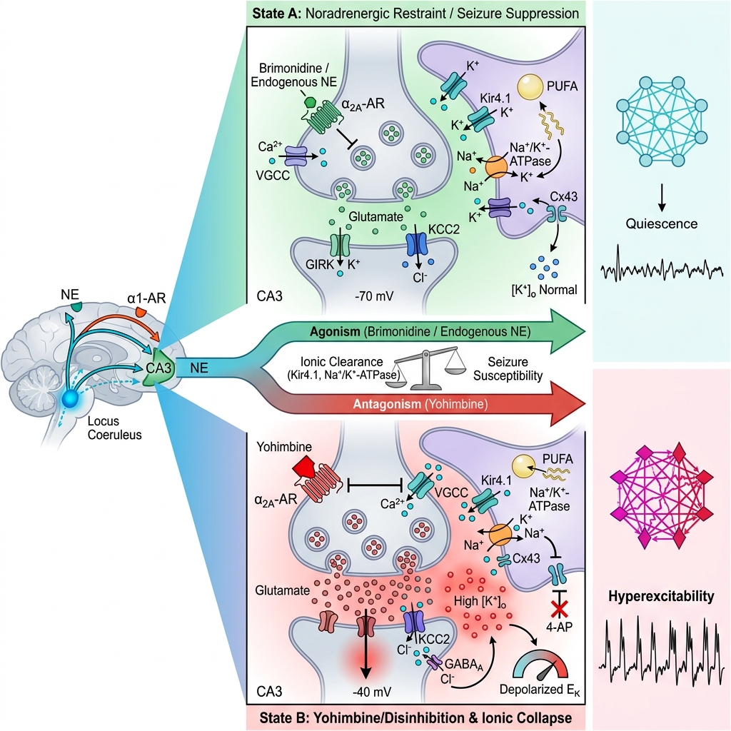
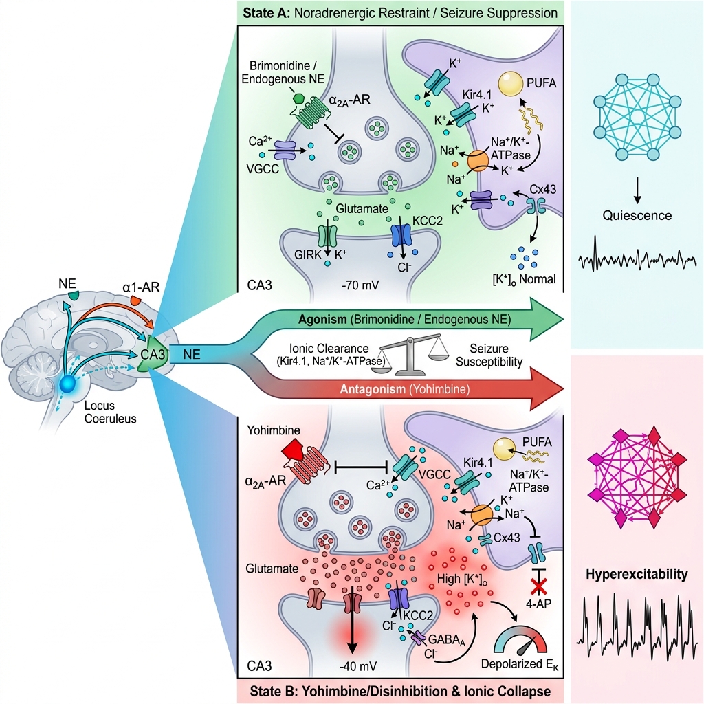
Graphical abstract
Introduction
Epilepsy is a chronic neurological disorder characterized by spontaneous, recurrent seizures arising from paroxysmal, highly synchronized neuronal discharges. Globally, epilepsy affects more than 50 million individuals, contributing substantially to the worldwide disease burden, with a disproportionately high prevalence in low- and middle-income countries1. The etiology of epilepsy is remarkably heterogeneous, encompassing genetic channelopathies, structural malformations, cerebral ischemia, central nervous system infections, and traumatic brain injury2. Beyond physical morbidity and seizure-related trauma, recurrent ictal events induce progressive cognitive impairment, psychiatric comorbidities, and diminished quality of life, underscoring the urgent imperative for more efficacious therapeutic paradigms3.
At the cellular and microcircuit level, epileptiform activity is conventionally conceptualized as an uncoupling of the balance between excitatory and inhibitory (E/I) synaptic transmission. Pathological network hypersynchrony is typically initiated by excessive glutamatergic excitation or compromised γ-aminobutyric acid (GABA)-mediated synaptic inhibition4,5. However, this classical synaptic doctrine is fundamentally grounded in the integrity of local transmembrane ionic gradients. In chronic epilepsy, ionic homeostasis is profoundly disturbed across the cerebral cortex and hippocampus, with marked impairment of astrocytic potassium buffering. This ionic dysregulation progressively degrades the electrochemical driving forces required for hyperpolarizing GABAergic transmission, thereby reinforcing network hyperexcitability and lowering seizure thresholds6,7.
The majority of conventional antiseizure medications (ASMs) were developed to restore synaptic E/I balance by potentiating GABAergic neurotransmission, blocking ionotropic glutamate receptors, or modulating voltage-gated sodium channels8. Despite the introduction of numerous third-generation ASMs over the past two decades, approximately one-third of patients remain pharmacoresistant, experiencing intractable, refractory seizures9. This persistent therapeutic impasse exposes the intrinsic limitations of targeting classical synaptic receptors and voltage-dependent conductances in isolation. Consequently, there is an imperative to explore alternative neuromodulatory systems capable of governing intrinsic network excitability and preserving the extracellular ionic microenvironment.
Despite recognizing the shortcomings of purely synaptic interventions, previous reviews have struggled to bridge conventional receptor pharmacology with non-synaptic glial biophysics10. Historically, neuromodulatory research has remained compartmentalized, prioritizing ligand-binding kinetics and classic synaptic inhibition while neglecting the overarching influence of the extracellular ionic milieu7. In particular, the dynamic fluctuations of extracellular potassium ([K]) during evoked epileptiform bursts are rarely integrated into pharmacological frameworks, leaving a critical knowledge gap regarding how ionic instability modulates—and is modulated by—endogenous neurochemical tone.
The central noradrenergic system is a powerful endogenous regulator of cerebral excitability. Norepinephrine (NE), acting through a diverse repertoire of G-protein-coupled adrenergic receptors (GPCRs), exerts profound modulatory control over neuronal firing rates, synaptic plasticity, and neurotransmitter release probability11. Preclinical investigations consistently demonstrate that noradrenergic signalling exerts robust anticonvulsant effects against diverse seizure-inducing stimuli12. In particular, the α-adrenergic receptor (α-AR) subtype represents a highly promising, non-canonical therapeutic target for suppressing epileptiform synchrony13,14.
Historically, investigations evaluating the noradrenergic modulation of seizure activity have focused almost exclusively on therapeutic agonism. Exogenous activation of α-ARs, using selective agonists such as brimonidine (UK14,304), is well documented to attenuate epileptiform discharges through presynaptic suppression of glutamate release and postsynaptic membrane hyperpolarization10. However, evaluating pharmacological suppression in isolation yields an incomplete understanding of network resilience. To comprehensively delineate the protective role of the noradrenergic system, a bidirectional pharmacological approach is indispensable. Employing a selective antagonist, such as yohimbine, serves as a vital counter-probe; by pharmacologically dismantling baseline noradrenergic tone, it unmasks the central nervous system's active dependence on endogenous norepinephrine to avert runaway hypersynchrony13.
While recent work by our group highlighted the therapeutic utility of selective α-AR agonism via agents such as brimonidine14, the present scoping review substantially broadens this paradigm by conceptualizing α-adrenergic signalling as a bidirectional governor of network excitability and extracellular potassium ([K]) dynamics. By unifying α-AR agonism with antagonist-mediated receptor blockade, this review delineates how endogenous noradrenergic tone coordinates seizure restraint, and how its therapeutic efficacy is fundamentally constrained by astrocytic K buffering capacity. Furthermore, this bidirectional lens illuminates the biophysical limitations of broad-spectrum potassium channel blockers (e.g., 4-aminopyridine) in ex vivo seizure models, providing a refined mechanistic foundation for evaluating α-AR therapeutics in pharmacoresistant epilepsy.
Methods
To systematically map the evidence linking receptor-mediated noradrenergic mechanisms to dynamic ionic homeostasis, we conducted a scoping review of the preclinical literature. The search strategy, screening, and data extraction were executed in strict accordance with the Preferred Reporting Items for Systematic Reviews and Meta-Analyses extension for Scoping Reviews (PRISMA-ScR) guidelines15.
Population–Concept–Context (PCC) Framework
To systematically structure the review objectives and define transparent eligibility criteria regarding the biophysical and pharmacological mechanisms of noradrenergic antiepileptic action, the scoping query was formulated using the Population–Concept–Context (PCC) framework (see Table 1 in the Tables and Figures section following the References).
Population–Concept–Context (PCC) Framework Defining Eligibility and Selection Criteria for Preclinical Studies.
| PCC Element | Inclusion Criteria | Exclusion Criteria |
|---|---|---|
| Population |
|
Studies exclusively utilizing non-neural peripheral tissues (e.g., cardiovascular, pulmonary, or renal models). Clinical human trials lacking translational mechanistic arms. |
| Concept |
Administration of norepinephrine or selective α2A-AR agonists (e.g., UK14,304 / brimonidine) and antagonists (e.g., yohimbine, atipamezole). Characterization of α2A-AR downstream effectors (e.g., GIRK channels, voltage-gated Ca2+ channels, cAMP/PKA signalling). Direct or inferred measurement of extracellular K+ dynamics, astrocytic spatial buffering (Kir4.1), or Na+/K+-ATPase activity. |
Pharmacological interventions exclusively targeting α1-, β1-, or β2-adrenergic receptors without evaluating α2A-ARs. Studies lacking characterization of either synaptic E/I balance or ionic/potassium biophysics. |
| Context |
Pathological states of epileptiform synchronization, hypersynchrony, or ictogenesis. Pharmacoresistant epilepsy paradigms. Disease models featuring disrupted ionic homeostasis, impaired astrocytic buffering, or cellular hyperexcitability (e.g., 4-aminopyridine, bicuculline, pilocarpine). |
Physiological neurocognitive studies (e.g., spatial learning, memory consolidation, sleep architecture) without an epilepsy or hyperexcitability component. Psychiatric models (e.g., depression, PTSD) lacking seizure susceptibility assessments. |
Population (P): Preclinical Epilepsy Models. The review targets biological models utilized to investigate epileptiform activity and seizure genesis. This encompasses ex vivo mammalian preparations (such as acute hippocampal or corticohippocampal slices exhibiting disrupted ionic buffering) and in vivo animal models of chemically, electrically, or genetically induced seizures. Clinical human trials and peripheral non-neural preparations were excluded to maintain focus on central network hyperexcitability.
Concept (C): α-Adrenergic Receptor Signalling and Potassium Dynamics. The core concept evaluates the neuromodulatory influence of central noradrenergic transmission, specifically focusing on the pharmacology of α-ARs using endogenous norepinephrine, selective agonists (e.g., brimonidine / UK14,304), and selective antagonists (e.g., yohimbine, atipamezole). The review evaluates how bidirectional α-adrenergic modulation intersects with two fundamental neurobiological mechanisms: (1) the pre- and postsynaptic regulation of the synaptic E/I balance, and (2) the biophysics of extracellular potassium ([K]) dynamics, inward-rectifier potassium (Kir4.1/GIRK) conductances, and astrocytic buffering capacity.
Context (C): Epileptogenesis and Ionic Dysregulation. The contextual setting is defined by pathological states of neuronal hypersynchrony, seizure propagation, and pharmacoresistance. The scope is restricted to studies examining paroxysmal discharges and disrupted ionic homeostasis in models where physiological network restraint has collapsed (e.g., 4-aminopyridine, bicuculline, or pilocarpine paradigms).
Search Strategy
A comprehensive systematic literature search was executed across three primary electronic databases: PubMed/MEDLINE, Scopus, and Web of Science. The final search was updated through June 2026. Search algorithms were constructed using combinations of Medical Subject Headings (MeSH) and title/abstract keywords coupled with Boolean operators. The explicit search strings were:
-
PubMed/MEDLINE: ("Receptors, Adrenergic, alpha-2"[MeSH] OR "Norepinephrine"[MeSH] OR "brimonidine" OR "yohimbine") AND ("Epilepsy"[MeSH] OR "Seizures"[MeSH] OR "epileptiform activity") AND ("Potassium"[MeSH] OR "potassium dynamics" OR "GIRK channels" OR "Astrocytes"[MeSH]) [Returned 45 records]
-
Scopus: TITLE-ABS-KEY(("alpha-2A adrenergic receptor" OR "norepinephrine" OR "brimonidine" OR "yohimbine") AND ("epilepsy" OR "seizures" OR "epileptiform activity") AND ("potassium dynamics" OR "GIRK channels" OR "astrocytes")) [Returned 38 records]
-
Web of Science: TS=(("alpha-2A adrenergic receptor" OR "norepinephrine" OR "brimonidine" OR "yohimbine") AND ("epilepsy" OR "seizures" OR "epileptiform activity") AND ("potassium dynamics" OR "GIRK channels" OR "astrocytes")) [Returned 22 records]
Language filters were applied to restrict inclusion to English-language peer-reviewed original research articles. To ensure comprehensive coverage, the reference lists of key reviews and authoritative original articles were hand-searched to identify additional eligible studies.
Eligibility Criteria
Studies were evaluated against predefined inclusion and exclusion criteria established in accordance with the PCC framework:
Inclusion Criteria: (1) Peer-reviewed original research articles; (2) preclinical in vivo, ex vivo, or in vitro biological models; (3) studies explicitly investigating the modulatory role of α-AR signalling, norepinephrine, selective α-AR agonists, or selective α-AR antagonists in seizure susceptibility or epileptiform activity; and (4) studies reporting mechanistic outcomes concerning synaptic transmission, ionic conductances, or extracellular potassium regulation.
Exclusion Criteria: (1) Studies strictly investigating peripheral adrenergic actions outside the central nervous system; (2) studies exclusively targeting α- or β-adrenergic receptors without characterizing α-ARs; (3) conference abstracts lacking full methodological datasets, narrative opinion pieces, and editorials; and (4) purely clinical observational studies lacking translational or biophysical validation.
Methodological Note on Preprints: To capture emerging mechanistic findings, preprints were considered eligible for preliminary screening when meeting PCC criteria; however, because preprints have not undergone formal peer review, their data were interpreted with methodological prudence.
PRISMA-ScR Flow Diagram
All identified records were imported into reference management software, and duplicate citations were removed. Two reviewers independently screened titles and abstracts against the eligibility criteria. Full-text articles of potentially eligible studies were subsequently retrieved and subjected to comprehensive evaluation. Disagreements regarding eligibility were resolved through discussion and consensus. The complete screening and selection cascade, detailing numbers identified, deduplicated, screened, excluded, and ultimately synthesized, is visually presented in Figure 1.
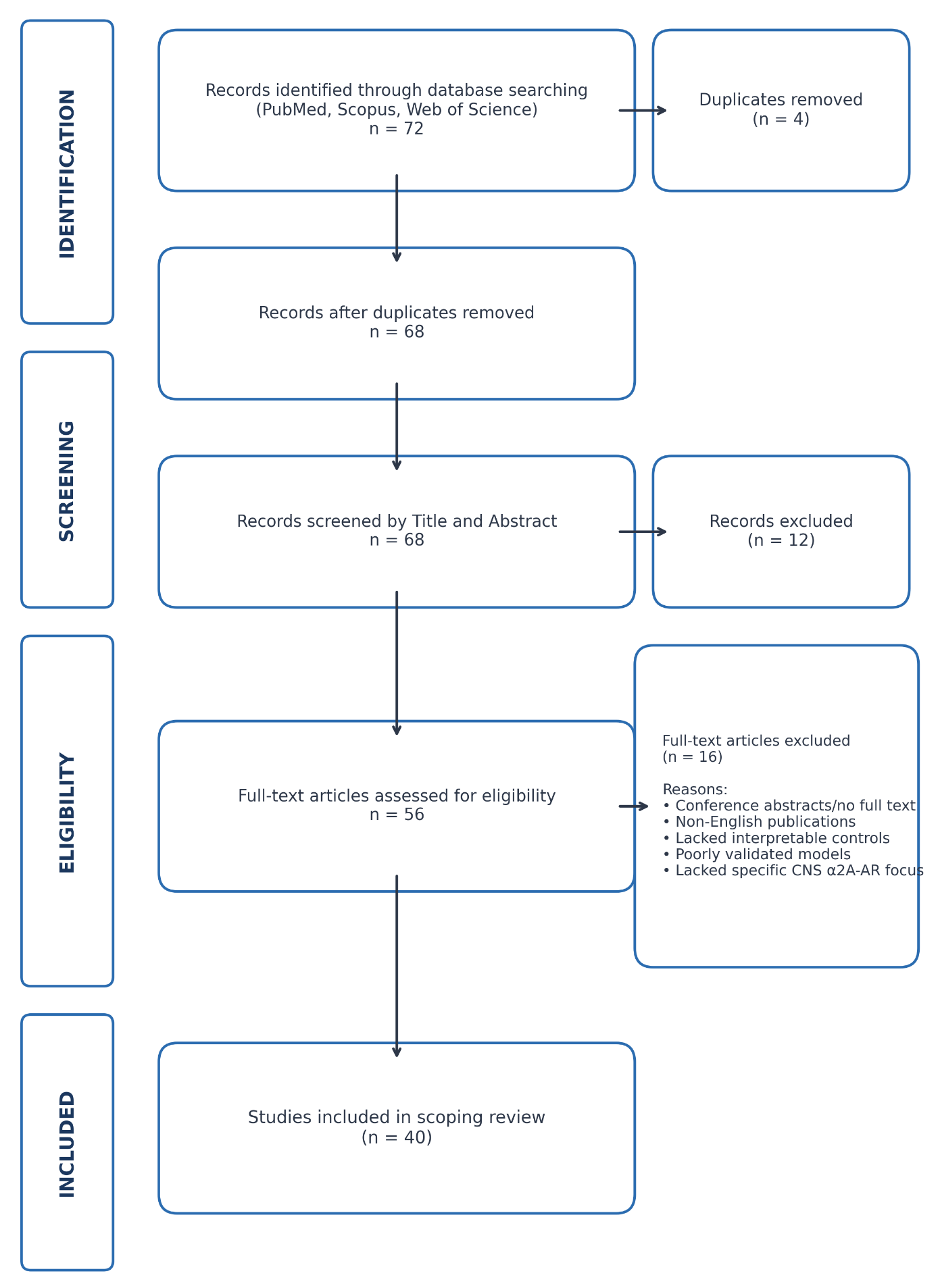
PRISMA-ScR Flow Diagram of Literature Search, Screening, and Study Selection Process. Systematic review workflow mapping the identification, deduplication, screening, eligibility appraisal, and final inclusion of preclinical literature in accordance with the Preferred Reporting Items for Systematic Reviews and Meta-Analyses extension for Scoping Reviews (PRISMA-ScR) guidelines. A systematic query executed across PubMed/MEDLINE (n = 45), Scopus (n = 38), and Web of Science (n = 22) yielded an initial total of 72 records. Following the removal of 4 duplicate records, 68 unique records were screened by title and abstract, resulting in the exclusion of 12 non-relevant articles. The remaining 56 full-text articles were rigorously evaluated against predefined Population–Concept–Context (PCC) eligibility criteria. Sixteen full-text articles were excluded for specific methodological reasons: conference abstracts lacking complete empirical datasets (n = 4), non-English publications (n = 3), absence of interpretable vehicle or control conditions (n = 3), poorly validated hyperexcitability models (n = 3), and lack of specific central nervous system α2A-AR focus (n = 3). A total of 40 primary research articles satisfied all inclusion criteria and were incorporated into the final qualitative evidence synthesis. Abbreviations: α2A-AR, α2A-adrenergic receptor; CNS, central nervous system; n, number of records/studies; PCC, Population–Concept–Context; PRISMA-ScR, Preferred Reporting Items for Systematic Reviews and Meta-Analyses extension for Scoping Reviews.
Data Extraction and Synthesis
A standardized data extraction form was developed to chart key study characteristics, including animal species, experimental preparation (in vivo vs. ex vivo), convulsant induction method, specific pharmacological agents administered, target receptor subtypes and ion channels, and primary neurophysiological outcomes. The full dataset of all 40 included studies was compiled into Supplementary Table S1. To provide an immediate mechanistic overview, a curated extraction of 13 representative studies detailing noradrenergic modulation, potassium biophysics, and astrocytic function is presented in Table 2. Direct institutional animal ethics approval was not required for this scoping review of published literature; nevertheless, all included primary studies were verified to have complied with national and institutional animal welfare guidelines (such as IACUC approval) as stated in their original publications.
Summary of Methodological Characteristics, Pharmacological Interventions, and Principal Findings from Representative Preclinical Studies on Noradrenergic Signalling and Potassium Biophysics in Epilepsy.
| S/N | Study Citation | Experimental Model & Preparation | Intervention & Target | Principal Preclinical Findings |
|---|---|---|---|---|
| 1 | Avoli et al., 2002 | 4-Aminopyridine (4-AP) and chemoconvulsants; targeting voltage-gated K+ channels and synaptic transmission. |
Demonstrated that 4-AP produces distinct epileptiform patterns: interictal discharges originating in CA3 and prolonged ictal-like discharges in the entorhinal cortex. Identified the "GABAergic paradox," wherein intense GABA release synchronizes pyramidal cells due to intracellular Cl- and extracellular K+ accumulation. Established that recurrent excitatory loops between hippocampus and parahippocampal cortices are required for seizure propagation. | |
| 2 | Jurgens et al., 2005 | Norepinephrine (NE) and subtype-selective adrenergic agonists and antagonists (α- and β-ARs). |
Demonstrated that exogenous NE robustly suppresses spontaneous epileptiform burst discharges in the CA3 subfield. Established that this anticonvulsant action is mediated by α2-ARs, as α2-agonists mimicked inhibition whereas α2-antagonists reversed NE actions. Revealed that β-AR activation exerts opposing proconvulsant effects, demonstrating subtype-dependent control of network excitability. | |
| 3 | Jurgens et al., 2007 | Brimonidine (UK14,304) and subtype-specific antagonists; targeting α2A-ARs. |
Confirmed that the selective α2A-AR agonist UK14,304 potently suppresses spontaneous CA3 burst discharges. Demonstrated subtype specificity using atipamezole and calculated pharmacological dissociation constants (Kb). Established that α2A-AR activation hyperpolarizes CA3 pyramidal neurons, providing an intrinsic brake against synchronized burst propagation. | |
| 4 | Kaila et al., 2014 | Mammalian neuronal networks and | Modulation of the KCC2 chloride cotransporter and extracellular ionic homeostasis. |
Showed that activity-induced elevations in extracellular potassium ([K+]o) impair KCC2-mediated chloride extrusion. Established the concept of "ionic plasticity," where depolarizing shifts in EGABA convert GABAergic inhibition into paradoxical excitation. Concluded that ionic collapse is a primary driver of network hyperexcitability and pharmacoresistance to GABAergic ASMs. |
| 5 | Ibrahim et al., 2021 | α2-AR antagonism (yohimbine) challenged with omega-3 fatty acids, diazepam, or combination. |
Systemic yohimbine alone rapidly induced clonic seizures (mean latency ~5 min) by eliminating endogenous noradrenergic restraint. Omega-3 fatty acid monotherapy exerted non-significant protection against yohimbine-provoked seizures. Combining omega-3 fatty acids with diazepam significantly delayed seizure onset and reduced seizure frequency compared to yohimbine alone. | |
| 6 | Galic et al., 2004 | Acute swim stress combined with selective receptor antagonists (α2-AR [yohimbine], glucocorticoid [mifepristone], opioid [naloxone]). |
Acute swim stress prior to pilocarpine administration delayed motor seizure onset via endogenous norepinephrine release. Pretreatment with yohimbine (5 mg/kg) completely abolished stress-induced anticonvulsant protection. Glucocorticoid and opioid receptor blockades failed to alter seizure latency, proving that stress protection depends specifically on α2-AR activation. | |
| 7 | Li et al., 2024 | Norepinephrine (NE); targeting free fatty acid (FFA) release and astrocytic Na+/K+-ATPase. |
Demonstrated that NE stimulates rapid polyunsaturated FFA release from astrocytic and neuronal membranes. Established that FFAs act as metabolic modulators that stimulate Na+/K+-ATPase pump activity. Concluded that NE-induced FFA production accelerates ion clearance, suppressing epileptiform bursts and terminating seizure discharges. | |
| 8 | Tsentsevitsky et al., 2022 | Modulation of GIRK channels and L-type voltage-gated Ca2+ channels. |
Demonstrated that GIRK channels serve as versatile regulators of transmitter exocytosis. Established that GIRK activity modulates calcium influx mediated by L-type Ca2+ channels. Concluded that by inhibiting Ca2+ entry, GIRK channels attenuate neurotransmitter release, linking K+ conductance to synaptic output. | |
| 9 | Niitani et al., 2023 | Norepinephrine and α1-adrenoceptor activation; seizure facilitation. |
Demonstrated that under compromised GABAergic inhibition, NE facilitates epileptiform discharges in mPFC layer 5 pyramidal neurons. Showed that this localized proconvulsant effect is mediated by α1-AR activation enhancing glutamatergic EPSCs via AMPA and NMDA receptors. | |
| 10 | Kiani et al., 2026 | Dorsoventral axis and circadian (night–day) regulation; extracellular K+ dynamics. |
Evaluated astrocytic [K+]ext dynamics across the longitudinal hippocampal axis and circadian cycles. Revealed that evoked network activity produces higher transient [K+]ext in the ventral hippocampus compared to the dorsal pole. Highlighted specialized regional and temporal dynamics in astrocytic ionic regulation, demonstrating region-specific vulnerability. | |
| 11 | Delling et al., 2002 | Neural cell adhesion molecule (NCAM) and lipid rafts; GIRK channel surface trafficking. |
Demonstrated that NCAM directs the cell-surface delivery and clustering of GIRK channels via lipid rafts. Established that membrane microdomain localization is required for functional G-protein-mediated K+ conductances and neuronal inhibition. | |
| 12 | Jin & Lu, 1998 | Novel peptide inhibitor (tertiapin); inward-rectifier K+ channels. |
Characterized tertiapin as a high-affinity peptide inhibitor that selectively blocks inward-rectifier potassium channels (Kir1.1, Kir3.1/3.4). Provided a critical pharmacological tool to isolate Kir and GIRK conductances in physiological and pathological studies. | |
| 13 | David et al., 2009 | Serum albumin exposure and TGF-β signalling; astrocytic Kir4.1 and GLT-1 regulation. |
Demonstrated that serum albumin exposure following blood–brain barrier breakdown activates astrocytic TGF-β signalling. Established that this pathway downregulates Kir4.1 potassium channels and GLT-1 transporters, impairing spatial K+ buffering and driving epileptogenesis. |
The Noradrenergic System in the Central Nervous System
Norepinephrine is synthesized from the amino acid L-tyrosine via a sequential enzymatic pathway within central and peripheral noradrenergic neurons16. In the mammalian central nervous system, the locus coeruleus (LC), a compact pontine nucleus located in the dorsal pons, constitutes the primary source of noradrenergic projections17. Ascending LC efferents diffuse widely throughout the entire neuroaxis, establishing dense, highly branched arborizations within excitable limbic structures, particularly the hippocampus and amygdala18. This extensive innervation positions the LC–NE axis as an endogenous master governor of network excitability, vigilance states, and seizure thresholds throughout the limbic system (see Graphical Abstract in the Tables and Figures section).
Norepinephrine exerts its diverse physiological actions by engaging adrenergic receptors, all of which belong to the superfamily of 7-transmembrane G-protein-coupled receptors (GPCRs)19. These receptors are phylogenetically and pharmacologically classified into two principal families: α and β. The α family is further subdivided into α (α, α, α) and α (α, α, α) subfamilies, whereas the β family comprises β, β, and β subtypes20. Agonist binding induces receptor conformational transitions that catalyze guanine nucleotide exchange on distinct heterotrimeric G-protein complexes. Through these distinct receptor subfamilies, noradrenergic signalling finely balances neural excitability and inhibition, dictating how networks respond to environmental stimuli and paroxysmal challenges21. Depending on the specific G-protein coupled to the receptor, downstream effector pathways can either hyperpolarize or depolarize target neurons, dynamically modulating cellular excitability22.
Among adrenergic receptor subfamilies, the α-AR class is uniquely configured to restrain neuronal hyperexcitability. By coupling preferentially to pertussis toxin-sensitive inhibitory G-proteins (G), α-AR activation inhibits adenylyl cyclase, lowers intracellular cyclic adenosine monophosphate (cAMP) accumulation, and suppresses voltage-gated calcium conductances23. This inhibitory cascade enables α-ARs to function as a powerful presynaptic "brake" on neurotransmitter release, suppressing excessive glutamate exocytosis during pathological synchronization while preserving baseline physiological synaptic transmission24,25.
Within this inhibitory class, the α-AR subtype is abundantly expressed across the mammalian brain and represents the primary mediator of noradrenergic inhibition. In the hippocampal formation, immunohistochemical and in situ hybridization studies localize α-ARs prominently across the strata oriens, radiatum, and lacunosum-moleculare of the CA1 and CA3 fields, as well as the dentate gyrus molecular layer26. Ultrastructurally, α-ARs are concentrated presynaptically on the axon terminals of both catecholaminergic and non-catecholaminergic neurons, functioning as auto- and heteroreceptors, respectively. Additionally, α-ARs are expressed postsynaptically on the dendritic shafts and spines of principal pyramidal neurons and are present on subpopulations of perisynaptic astrocytes23,26.
Signal transduction via α-ARs is tightly coupled to Gα subunits. Receptor activation drives the direct inhibition of adenylyl cyclase, reducing intracellular cAMP synthesis and attenuating protein kinase A (PKA) activity23. Concurrently, liberated Gβγ dimers interact directly with membrane ion channels, restricting presynaptic calcium entry while promoting postsynaptic potassium efflux. Together, these coordinated actions establish α-ARs as pivotal biophysical dampers of circuit hyperexcitability.
α-AR Modulation of Synaptic Transmission
Presynaptic Mechanisms
Activation of presynaptic α-ARs potently attenuates neurotransmitter release by modulating vesicular exocytosis and terminal calcium influx. Following agonist binding, the dissociated Gβγ dimer directly binds to presynaptic voltage-gated calcium channels (VGCCs)—predominantly N-type (Ca2.2) and P/Q-type (Ca2.1) channels—producing a depolarizing voltage shift in channel activation that limits calcium entry during action potential arrival27,28. Concurrently, the Gα subunit inhibits adenylyl cyclase, suppressing cAMP-dependent PKA activity23. Because PKA normally phosphorylates key components of the SNARE complex and active zone proteins (such as synapsin and rabphilin), its downregulation uncouples action potential arrival from the exocytotic machinery. Through this dual mechanism of restricted calcium influx and impaired vesicle fusion, presynaptic α-AR activation curtails the release of classical transmitters, including glutamate and γ-aminobutyric acid (GABA)29.
Postsynaptic Mechanisms
Beyond presynaptic modulation, α-AR signalling reduces postsynaptic neuronal excitability through direct membrane hyperpolarization. This inhibitory effect is mediated primarily by G-protein-activated inwardly rectifying potassium (GIRK / Kir3) channels30. Agonist binding to postsynaptic α-ARs liberates Gβγ subunits that bind directly to cytoplasmic domains of GIRK channel heterotetramers (GIRK1/GIRK2), promoting channel opening. Furthermore, in certain hyperexcitable microcircuits, presynaptic GIRK conductances modulate network tone: pharmacological blockade of presynaptic GIRK channels has been reported to relieve auto-inhibition and enhance GABA release from interneuron terminals, thereby reinforcing synaptic inhibition31.
The opening of postsynaptic GIRK channels drives a rapid potassium (K) efflux out of the intracellular compartment and into the extracellular space. This outward K current hyperpolarizes the neuronal membrane, shifting the resting membrane potential toward the potassium equilibrium potential (E) and away from the action potential threshold32,33. Consequently, postsynaptic α-AR activation exerts a robust biophysical dampening effect, reducing neuronal firing rates and suppressing repetitive burst generation.
The E/I Paradox
The simultaneous presynaptic suppression of both glutamate and GABA release creates a theoretical paradox: if α-AR activation dampens excitatory and inhibitory transmission concurrently, how does it reliably exert a net anticonvulsant effect? The resolution to this excitatory/inhibitory (E/I) paradox lies in the differential receptor sensitivity and circuit-specific distribution of α-ARs across hippocampal networks. First, presynaptic α-ARs exhibit differential pharmacological efficacy, suppressing excitatory glutamatergic terminals more robustly than inhibitory GABAergic terminals in key hippocampal pathways, thereby shifting the net synaptic drive toward inhibition34. Second, robust postsynaptic hyperpolarization driven by GIRK channel activation provides a dominant shunting conductance13. Even when GABA exocytosis is reduced, persistent postsynaptic hyperpolarization silences principal pyramidal cells, preventing the synchronization of burst discharges required for epileptiform propagation.
Crucially, this paradox is resolved by the anatomical distribution of α-ARs within the hippocampal microcircuit. In the CA3 subfield—a primary pacemaker for limbic seizures—presynaptic α-ARs are densely clustered on the recurrent axon collaterals of CA3 pyramidal neurons, but are expressed at significantly lower levels on local GABAergic basket interneurons13. Consequently, noradrenergic activation preferentially silences recurrent excitatory auto-amplification while sparing feedforward and feedback GABAergic inhibition12,13. This circuit-specific configuration ensures that the net network consequence of α-AR activation remains decisively inhibitory.
The Intersection of α Signalling and Extracellular K Dynamics
GIRK Channels and K Efflux
The α-adrenergic receptor attenuates network hyperexcitability via G-coupled pathways, with postsynaptic GIRK channel opening playing a prominent role. However, the efficacy of GIRK-mediated hyperpolarization depends on proper surface trafficking and microdomain localization of channel complexes. Membrane targeting of GIRK channels is coordinated by lipid rafts and regulated by neural cell adhesion molecules (NCAM), which facilitate channel clustering at postsynaptic densities30,35. Beyond direct electrophysiological shunting, α-AR activation suppresses adenylyl cyclase activity, leading to sustained reductions in intracellular cAMP levels36. Because elevated cAMP promotes PKA-mediated phosphorylation of glutamate receptors and hyperpolarization-activated pacemaker currents, suppression of this signalling cascade dampens long-term network hyperresponsiveness.
Furthermore, decreased cAMP signalling inhibits hyperpolarization-activated cyclic nucleotide-gated (HCN) channels, which conduct the pacemaker current (I) that supports repetitive rhythmic firing. Additionally, certain α-AR agonists, including guanabenz, have been shown to directly block HCN channel conductances37. Downregulation of I reduces intrinsic neuronal excitability and curtails the ability of pyramidal networks to sustain paroxysmal burst discharges.
The opening of GIRK channels drives potassium ions down their electrochemical gradient from the intracellular space into the confined extracellular space (ECS). Although this outward current hyperpolarizes the neuron, it imposes an activity-dependent K load onto the perisynaptic microenvironment. Consequently, the sustained efficacy of α-mediated hyperpolarization is tethered to the rate of extracellular potassium ([K]) clearance38. In healthy brain tissue, extracellular K is rapidly cleared by astrocytic homeostatic networks via inwardly rectifying Kir4.1 channels and active uptake by Na/K-ATPase pumps. However, this clearance capacity is not uniform throughout the brain. Marked dorsoventral variations exist across the hippocampus; recent electrophysiological evidence indicates that during evoked network activity, the ventral hippocampus experiences significantly greater transient increases in [K] than the dorsal hippocampus39. This observation reflects activity-dependent accumulation rather than inherent clearance failure, highlighting that the ventral pole is uniquely vulnerable to hyperexcitability if glial clearance mechanisms become overwhelmed39.
In pathological tissue where astrocytic K regulation fails—such as the downregulation of Kir4.1 channels observed in temporal lobe epilepsy40—clearance is severely compromised. Under these conditions, GIRK-mediated K efflux can exacerbate pathological elevations in [K]. As extracellular K accumulates, the transmembrane K gradient collapses, diminishing the driving force required for further hyperpolarization. Furthermore, unbuffered [K] accumulation depolarizes adjacent neurons, paradoxically amplifying the network hypersynchrony that α-AR activation was recruited to suppress41,42.
The therapeutic impact of GIRK channel activation is also circuit-dependent. In focal depolarizing pathologies like temporal lobe epilepsy, α-AR-induced GIRK activation provides robust anticonvulsant protection by suppressing hyperexcitable circuits. Conversely, in thalamocortical networks, excessive hyperpolarization can de-inactivate T-type Ca channels, facilitating rebound burst firing and absence seizure generation. In this context, anti-absence medications such as ethosuximide have been reported to modulate GIRK-related conductances alongside their canonical thalamic targets43. This circuit divergence demonstrates that the therapeutic utility of α-AR agonists must be evaluated within specific anatomical contexts, proving most effective in focal limbic epilepsies where persistent depolarization and impaired potassium buffering drive epileptogenesis.
Astrocytic K Clearance as a Network Anchor
The hyperpolarizing efficacy of GIRK channel activation is fundamentally dependent on the rapid clearance of extruded K from the extracellular space. Astrocytes serve as the principal biophysical anchors of this ionic equilibrium. Under physiological conditions, astrocytes clear excess [K] via two cooperative mechanisms: inward-rectifying Kir4.1 channels, which allow passive K influx into the glial syncytium, and active uptake mediated by astrocytic Na/K-ATPase pumps6. Once inside the syncytium, K is redistributed across extensive gap-junction networks (formed by connexins Cx43 and Cx30) to distant regions of lower concentration—a process known as spatial potassium buffering6. Pharmacological tools, such as the selective peptide inhibitor tertiapin, have helped characterize how Kir4.1 and GIRK conductances maintain this resting membrane stability44. Furthermore, in hyperexcitable networks, astrocytes contribute directly to adrenergic modulation: α-AR stimulation on astrocytes stimulates intracellular calcium transients that trigger the non-vesicular release of GABA, providing an astrocyte-derived source of synaptic inhibition45. This indicates that astrocytic adrenergic receptors may represent an auxiliary pathway for restoring inhibitory tone in diseased tissue.
However, during epileptogenesis, this astrocytic anchor undergoes severe structural and functional breakdown. In chronic temporal lobe epilepsy, reactive gliosis leads to profound downregulation and mislocalization of Kir4.1 channels40. Moreover, chronic epilepsy disrupts gap-junction coupling, causing uncoupling of the astrocytic syncytium46. Consequently, spatial potassium buffering is impaired throughout the sclerotic hippocampus40. When astrocytic buffering capacity is exceeded, activity-dependent K extrusion accumulates within the ECS41.
As [K] rises from its physiological baseline (~3 mM) to pathological levels (8–12 mM), the neuronal potassium equilibrium potential (E) depolarizes significantly according to the Nernst equation. This collapse of the electrochemical driving force neutralizes the hyperpolarizing current mediated by GIRK channels, rendering α-AR-mediated inhibition functionally ineffective30. Without an operative astrocytic K sink, the loss of hyperpolarizing drive strips the microcircuit of intrinsic protective mechanisms, lowering the threshold for paroxysmal depolarization shifts and spontaneous epileptiform bursts41,42.
The Chloride Gradient Collapse
Sustained elevations in [K] undermine the inhibitory framework of the brain by dismantling neuronal chloride homeostasis. Hyperpolarizing GABAergic transmission via ionotropic GABA receptors depends strictly on a low intracellular chloride concentration ([Cl]), which is actively maintained in mature neurons by the potassium-chloride cotransporter 2 (KCC2). KCC2 couples the extrusion of one Cl ion to the favorable outward gradient of one K ion. When [K] rises due to astrocytic clearance failure, the thermodynamic driving force for outward KCC2 cotransport is degraded, and the transporter can even run in reverse7.
This transport failure causes intracellular Cl accumulation, shifting the GABAergic reversal potential (E) to depolarized values above the neuronal resting membrane potential. Consequently, GABA receptor opening elicits chloride efflux rather than influx, converting GABAergic transmission from an inhibitory shunting mechanism into an excitatory, depolarizing drive. This phenomenon—termed "ionic plasticity"—fundamentally promotes network hyperexcitability and contributes to pharmacoresistance against conventional GABA-enhancing drugs47.
This pathological transition closely recapitulates early neurodevelopmental physiology. During embryogenesis and early postnatal development, immature neurons express low levels of KCC2 and high levels of the chloride-importing cotransporter NKCC1, resulting in elevated [Cl]. In this immature state, GABA is depolarizing and drives spontaneous network synchrony, known as giant depolarizing potentials (GDPs), which are essential for circuit maturation48. In chronic epilepsy, KCC2 downregulation and persistent K accumulation cause mature networks to regress to this depolarizing state49. This ionic shift synergizes with compromised astrocytic K buffering, generating a microenvironment permissive for autonomous epileptiform discharges.
In this context, the presynaptic efficacy of α-AR activation is vital. Although postsynaptic α-AR activation elicits modest K efflux through GIRK channels33, this local ionic load is dwarfed by the receptor's powerful presynaptic inhibition. By suppressing high-frequency glutamatergic firing, α-AR signalling prevents the massive, activity-dependent K efflux that accompanies prolonged ictal discharges. Thus, noradrenergic tone indirectly protects KCC2 transport and preserves hyperpolarizing GABAergic inhibition, acting as an essential upstream stabilizer of transmembrane chloride gradients7.
Noradrenergic Actions During Epileptiform Activity
The Dual Role of NE
The neuromodulatory impact of norepinephrine in epileptiform activity is functionally divergent. Depending on the targeted brain region, receptor subtype, and microcircuit architecture, NE exhibits both anticonvulsant and proconvulsant properties50. This functional duality is governed by the downstream G-protein coupling profiles of the adrenergic receptor families. While G-coupled α-ARs consistently inhibit network excitability, G-coupled α-ARs and G-coupled β-ARs stimulate phospholipase C and adenylyl cyclase cascades, respectively. In vulnerable networks, these pathways elevate intracellular calcium and cAMP, potentiating excitatory currents and promoting synchronization11.
Within the hippocampal formation, noradrenergic innervation functions as an endogenous brake on hyperexcitability. Dense LC afferents release NE to engage α-ARs, maintaining high seizure thresholds. Depleting central noradrenergic tone—either chemically via the selective neurotoxin DSP-4 or genetically in dopamine β-hydroxylase (DBH) knockout mice—significantly accelerates kindling progression and enhances susceptibility to chemical convulsants14,50. Conversely, pharmacological amplification of noradrenergic transmission or direct α-AR agonism raises seizure thresholds and attenuates epileptiform propagation.
However, this anticonvulsant profile is not universal. In certain neocortical and thalamocortical networks, noradrenergic stimulation can facilitate epileptiform discharges11,51. In deep neocortical layers, β-adrenergic receptor activation enhances persistent sodium currents (I) and amplifies L-type Ca conductances, triggering paroxysmal depolarizing shifts. Similarly, in thalamic relay neurons, noradrenergic depolarization can shift network firing patterns into oscillatory bursting, exacerbating spike-wave discharges in absence epilepsy models11,51. Thus, the net outcome of noradrenergic interventions depends on the receptor subtype targeted and the regional circuit anatomy.
Glutamatergic Promotion vs Astrocytic Protection
A striking example of regional divergence occurs in the medial prefrontal cortex (mPFC). In contrast to its protective role in the hippocampus, NE can promote epileptiform synchrony in the mPFC by engaging α- and β-adrenergic receptors11,52. In layer 5 mPFC pyramidal neurons, α-AR stimulation induces robust spontaneous excitatory postsynaptic currents (EPSCs) and enhances AMPA and NMDA receptor-mediated currents52,53. Under conditions of reduced GABAergic inhibition, noradrenergic activation exacerbates epileptiform discharge frequency and severity in prefrontal microcircuits53.
Conversely, robust noradrenergic signalling recruits astrocyte-mediated protective mechanisms to counteract network hyperexcitability. Recent findings demonstrate that acute NE exposure triggers the rapid release of polyunsaturated free fatty acids (FFAs) from neuronal and astrocytic plasma membranes54. These FFAs function as metabolic messengers that stimulate astrocytic Na/K-ATPase activity. By accelerating active K and Na reuptake, NE-induced FFA release enhances glial ionic buffering, accelerating the termination of seizure-like discharges and restoring microenvironmental stability54.
Systemic Neuromodulation and Autoreceptor Dynamics
In addition to local actions within epileptogenic foci, systemic administration of α-AR agonists alters global brain states by activating inhibitory autoreceptors. Genetic knockout studies confirm that the α-AR subtype functions as the primary autoreceptor mediating negative feedback on noradrenergic transmission55. In the locus coeruleus, clonidine activates somatodendritic α autoreceptors, hyperpolarizing LC neurons and suppressing spontaneous firing55,56. Concurrently, α autoreceptors located on axon terminals inhibit activity-dependent NE exocytosis throughout the neuroaxis56,57. In vivo microdialysis confirms that systemic α-AR agonism significantly reduces extracellular NE concentrations in the hippocampus58. This decrease in noradrenergic outflow dampens ascending subcortical arousal and diminishes generalized cortical excitability, providing a systemic mechanism that impedes seizure propagation.
Nevertheless, systemic α autoreceptor activation presents a major clinical bottleneck14. Because the LC–NE system regulates wakefulness, arousal, and autonomic function, non-selective systemic α agonists invariably cause severe sedation, bradycardia, and hypotension23,51. For example, dexmedetomidine suppresses LC firing and disinhibits the sleep-promoting ventrolateral preoptic nucleus (VLPO), inducing a state that electroencephalographically resembles non-rapid eye movement (NREM) sleep59. These adverse effects limit the long-term therapeutic utility of systemic agonists like clonidine in chronic epilepsy14. Consequently, translational drug development is shifting toward subtype-selective allosteric modulators, biased agonists, or targeted local drug-delivery strategies60,61 that selectively engage α-ARs within hyperexcitable limbic foci while sparing systemic autonomic and arousal circuitry.
Bidirectional Modulation of Network Excitability: The Opposing Dynamics of α-Adrenergic Agonism and Antagonism
The therapeutic potential of α-adrenergic modulation in epilepsy is best understood through a bidirectional framework rather than a binary on–off switch. Endogenous norepinephrine provides a constitutive level of α-AR activation that actively restrains runaway network synchronization62. Comparing pharmacological receptor activation with antagonist-mediated removal of endogenous tone reveals the degree to which baseline seizure thresholds depend on noradrenergic signalling.
Selective α-AR agonists, such as brimonidine (UK14,304), demonstrate the therapeutic efficacy of reinforcing this inhibitory axis, suppressing epileptiform bursts in hyperexcitable networks without disrupting baseline neurotransmission13. Conversely, the vital contribution of endogenous α-AR tone is revealed when this pathway is pharmacologically blocked. Yohimbine, a competitive α-AR antagonist, eliminates this endogenous brake, unmasking latent hyperexcitability, lowering seizure thresholds, and accelerating seizure progression in preclinical models63. The following subsections examine these opposing pharmacological strategies in detail.
Brimonidine (UK14,304): Pharmacological Profile and Experimental Caveats
Selectivity and Neuroprotection
Brimonidine (UK14,304) is a selective, high-affinity α-AR agonist belonging to the 5-bromoquinoxaline derivative family64. It displays a high binding selectivity for the α subtype over α- and β-adrenergic receptors (selectivity ratio >1000:1), minimizing off-target adverse effects65. By mimicking endogenous norepinephrine, UK14,304 inhibits presynaptic glutamate release and drives postsynaptic hyperpolarization66. In addition to suppressing epileptiform discharges, brimonidine exerts neuroprotective effects against glutamate-mediated excitotoxicity in hippocampal and retinal models67. Consequently, brimonidine represents an optimal pharmacological tool for isolating the antiepileptic actions of α-AR signalling.
Re-evaluating Ex Vivo Efficacy (The 4-AP Caveat)
Although brimonidine provides robust neuroprotection, its reported anticonvulsant efficacy in certain ex vivo electrophysiological models—particularly the 4-aminopyridine (4-AP) hippocampal slice model—appears modest. In acute hippocampal slices, brimonidine does not alter baseline field excitatory postsynaptic potentials (fEPSPs) evoked by Schaffer collateral stimulation in CA1 stratum radiatum12. In the presence of 4-AP, brimonidine reduces the frequency of spontaneous interictal-like events (IIEs), significantly prolonging the inter-event interval, but does not alter discharge amplitude12.
Interpreting this modest efficacy requires evaluating the network architecture of the 4-AP model. Spontaneous epileptiform discharges in 4-AP-treated slices originate within the recurrently connected CA3 pyramidal network before propagating along Schaffer collaterals to the CA1 field68. Foundational studies demonstrate that α-AR activation potently suppresses epileptiform burst discharges in the CA3 subfield13. Thus, the reduction in interictal event frequency recorded downstream in CA1 reflects α-AR-mediated suppression of the primary CA3 pacemaker, rather than local CA1 inhibition12,13.
Biophysical Model Limitations
The moderate efficacy of brimonidine observed in 4-AP models is primarily an artefact of the model's pharmacological mechanism rather than an intrinsic limitation of α-AR therapy. 4-AP is a broad-spectrum blocker of voltage-gated potassium channels (predominantly K1.1–1.4 and K3.1), which are responsible for fast repolarization and A-type currents68. Crucially, the downstream inhibitory actions of α-AR signalling rely heavily on potassium conductances, including GIRK channels. By pharmacologically blocking K channels with high concentrations of 4-AP (typically 50–100 μM), the model directly impairs the potassium conductances required for α-AR-mediated hyperpolarization.
Furthermore, blocking delayed rectifier and transient K channels broadens presynaptic action potentials, triggering massive calcium influx and unconstrained neurotransmitter release. The resulting paroxysmal activity drives rapid and pathological [K] accumulation in the restricted extracellular space42,69. As [K] rises, the local potassium equilibrium potential (E) depolarizes, degrading the driving force for GIRK-mediated hyperpolarization7,30. Therefore, testing an α-AR agonist in a model that paralyzes potassium conductances blunts the drug's primary mechanism of action. This biophysical limitation highlights the need to validate α-AR modulators in spontaneous epilepsy models with intact ionic conductances.
Yohimbine as a Pharmacological Counter-Probe: Mechanistic Insights and Experimental Limitations
Yohimbine is an indole alkaloid that acts as a competitive antagonist of α-adrenergic receptors, functioning systemically as a sympathomimetic agent that increases central and peripheral noradrenergic release70,71. While selective agonists demonstrate the therapeutic benefit of receptor stimulation, antagonist counter-probes like yohimbine unmask the vital role of endogenous noradrenergic tone in maintaining network stability24.
By competitively blocking presynaptic α-ARs, yohimbine removes baseline auto- and heteroreceptor inhibition on glutamatergic terminals. The resulting disinhibition leads to unchecked glutamate release and excessive pyramidal cell firing. This hyperactive state accelerates activity-dependent [K] accumulation in the perisynaptic space72. Yohimbine accelerates the rate at which neuronal firing exceeds astrocytic K clearance capacity. This effect illustrates how rapidly unchecked excitation can overwhelm glial buffering networks.
However, the consequences of α-AR antagonism are circuit-specific. In recurrent limbic networks like the hippocampal CA3 subfield, yohimbine exacerbates epileptiform bursting by removing baseline noradrenergic restraint. In contrast, in thalamocortical circuits responsible for absence seizures, α antagonists can suppress spike-wave discharges by altering thalamic relay neuron polarization73. Thus, yohimbine does not inherently generate hyperexcitability; rather, it accelerates the transition to seizure thresholds by removing endogenous inhibition. This highlights that the protective efficacy of the noradrenergic system is interdependent with astrocytic K buffering capacity.
Experimental Validation: Yohimbine in Preclinical Seizure Models
In vivo preclinical studies confirm that yohimbine possesses potent proconvulsant properties, providing direct evidence of endogenous noradrenergic protection. In the lithium-pilocarpine model, acute swim stress induces robust endogenous norepinephrine release, which significantly delays the onset of motor seizures. However, pretreatment with yohimbine (5 mg/kg) completely abolishes this stress-induced neuroprotection, accelerating seizure onset to non-stressed baseline levels74.
Ex vivo slice electrophysiology further substantiates these findings. Initial investigations in rat hippocampal slices demonstrated that non-selective α antagonists, including yohimbine, eliminate the endogenous noradrenergic suppression of CA3 epileptiform bursting12. Subsequent work using selective antagonists (atipamezole) and agonists (brimonidine) determined apparent equilibrium dissociation constants (K), confirming that the α-AR subtype is the precise receptor mediating this endogenous inhibition13.
At high systemic doses (e.g., 40–45 mg/kg in rodents), yohimbine reliably induces clonic seizures75. By removing the presynaptic noradrenergic brake, yohimbine triggers massive glutamatergic discharge that rapidly overwhelms astrocytic K clearance, causing severe [K] accumulation76,77. These yohimbine-induced seizure models provide valuable platforms for evaluating how excessive excitation breaches glial buffering capacity, precipitating microenvironmental collapse.
Together, the brimonidine and yohimbine data establish that α-ARs function as bidirectional regulators of network excitability. However, the translational utility of classical agonists and antagonists is restricted by systemic cardiovascular and sedative adverse effects11. Future clinical translation requires the development of circuit-specific and subtype-selective modulators capable of engaging focal α-ARs without triggering systemic autonomic disruption.
Limitations of the Review
Several methodological limitations of this scoping review should be considered. First, restricting the systematic search to PubMed/MEDLINE, Scopus, and Web of Science may have omitted relevant grey literature, unpublished negative findings, and institutional theses. Second, the synthesized evidence relies predominantly on rodent ex vivo slice preparations and acute preclinical seizure models. While these systems are optimal for dissecting receptor pharmacology and ionic biophysics, they lack the full systemic complexity of human epilepsy, including intact neurovascular units, neuroinflammatory cascades, and human-specific cytoarchitectural organization. Consequently, translating these mechanistic insights to clinical pharmacoresistant epilepsy requires rigorous validation in chronic disease models and human resected tissue.
Conclusion and Future Directions
In conclusion, evaluating antiseizure interventions requires considering the broader biophysical context of the epileptic microenvironment. Network excitability is governed not only by classical synaptic E/I balance, but also fundamentally by extracellular ionic homeostasis, astrocytic spatial buffering, and endogenous neuromodulatory tone. While α-AR activation dampens network hyperexcitability through presynaptic suppression of glutamate exocytosis and postsynaptic hyperpolarization via GIRK channels, these protective mechanisms are strictly dependent on intact extracellular potassium ([K]) regulation and functional astrocytic buffering.
When astrocytic K clearance fails, [K] accumulation collapses the transmembrane potassium gradient, inactivates KCC2 cotransport, shifts GABAergic signalling toward depolarization, and neutralizes α-AR-mediated hyperpolarization. As demonstrated by pharmacological counter-probes like yohimbine, endogenous noradrenergic tone actively restrains excessive excitation under physiological conditions. Evaluating α-AR pharmacology in models that artificially block potassium conductances (such as 4-aminopyridine) blunts the receptor's primary mechanisms of action, highlighting the need to test candidates in spontaneous seizure models with intact ionic conductances.
Future preclinical research should prioritize in vivo chronic epilepsy models that preserve glial networks, physiological ionic dynamics, and circadian rhythms. Integrating bidirectional receptor pharmacology with glial ionic homeostasis provides a comprehensive framework for developing novel antiseizure therapies, including chronotherapeutic strategies tailored to periods of heightened ionic vulnerability in pharmacoresistant epilepsy.
Declarations
Abbreviations
4-AP: 4-aminopyridine; AEDs: antiepileptic drugs (antiseizure medications); AMPA: α-amino-3-hydroxy-5-methyl-4-isoxazolepropionic acid; cAMP: cyclic adenosine monophosphate; Cl: chloride; CNS: central nervous system; DBH: dopamine β-hydroxylase; E/I: excitation/inhibition; E: GABAergic reversal potential; E: potassium reversal potential; EPSC: excitatory postsynaptic current; FFA: free fatty acid; GABA: γ-aminobutyric acid; GABA: γ-aminobutyric acid type A; GDP: giant depolarising potential; GIRK: G-protein-activated inwardly rectifying potassium channel; GLT-1: glutamate transporter 1; GPCR: G-protein-coupled receptor; HCN: hyperpolarisation-activated cyclic nucleotide-gated channel; IACUC: Institutional Animal Care and Use Committee; I: hyperpolarisation-activated pacemaker current; IIE: interictal-like event; I: persistent sodium current; K: potassium; [K]: extracellular potassium concentration; [K]: extracellular potassium concentration; KCC2: potassium-chloride cotransporter 2; Kir4.1: inwardly rectifying potassium channel subunit 4.1; LC: locus coeruleus; MeSH: Medical Subject Headings; mPFC: medial prefrontal cortex; NCAM: neural cell adhesion molecule; NE: norepinephrine; NMDA: N-methyl-D-aspartate; NMJ: neuromuscular junction; NREM: non-rapid eye movement; PCC: Population–Concept–Context; PKA: protein kinase A; PRISMA-ScR: Preferred Reporting Items for Systematic Reviews and Meta-Analyses extension for Scoping Reviews; PTDF: Petroleum Technology Development Fund; TGF-β: transforming growth factor-beta; VGCC: voltage-gated calcium channel; VLPO: ventrolateral preoptic nucleus; α-AR: α-adrenergic receptor.
Acknowledgments
We express our profound gratitude to Dr. Anton I. Ivanov and Dr. Christophe Bernard for their invaluable mentorship, scientific direction, and unwavering support throughout the conceptualisation of this work.
Author’s contributions
KA and DA conceptualised the study and designed the scoping review protocol. KA, DA, SMM, AAJ, AA, MMM, and BIM performed the literature search, study screening, data extraction, and quality appraisal. KA drafted the initial manuscript. DA, SMM, AAJ, AA, MMM, and BIM contributed to critical revisions and scientific refinement. All authors read and approved the final manuscript.
Funding
This work was supported by a doctoral scholarship awarded to Kabeer Abubakar by the Petroleum Technology Development Fund (PTDF), Nigeria. The funding body played no role in the design of the study, collection, analysis, or interpretation of data, or in writing the manuscript.
Availability of data and materials
All data generated or analysed during this scoping review are included in this published article, its tables, figures, and supplementary materials (Supplementary Table S1).
Ethics approval and consent to participate
Not applicable. This manuscript is a scoping review of previously published literature and did not involve primary research with human participants or live animals conducted directly by the authors. All included preclinical studies were verified to have received appropriate institutional animal care and ethics committee approvals as reported in their original publications.
Consent for publication
Not applicable.
Declaration of generative AI and AI-assisted technologies in the writing process
During the preparation of this manuscript, the authors utilised Gemini (Google) solely for language editing, stylistic refinement, and grammatical polishing, as well as to assist in conceptualising the layout of the graphical abstract. The authors reviewed and edited all generated text, take full responsibility for the scientific integrity and content of this publication, and confirm that no generative AI was used for data fabrication or ungrounded synthesis.
Competing interests
The authors declare that they have no competing interests (financial or non-financial).