
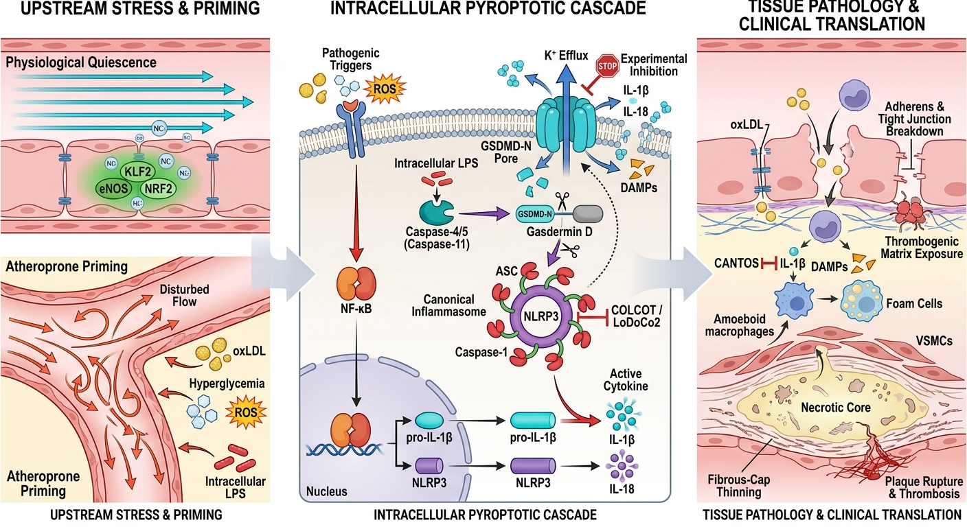


IL-1β-Centric Pyroptotic Signaling in Vascular Endothelium: A Missing Link in Cardiovascular Disease Progression
- Meenakshi Academy of Higher Education and Research (Deemed to be University), Chennai, Tamil Nadu, India
- Department of Anatomy, Meenakshi Medical College Hospital and Research Institute, Meenakshi Academy of Higher Education and Research (Deemed to be University), Kanchipuram, Tamil Nadu, India
- Department of Anatomy, Government Medical College Kannur, Pariyaram, Kerala, India
- Department of Microbiology, DMWIMS Medical College, Wayanad, Kerala, India
- Department of Cytogenetics, Genetika, Centre for Advanced Genetic Studies, Thiruvananthapuram, Kerala, India
Abstract
Background: Cardiovascular disease (CVD) remains the leading cause of morbidity and mortality worldwide, with chronic vascular inflammation serving as a primary driver of disease initiation, progression, and plaque destabilization. Accumulating evidence implicates endothelial pyroptosis as a critical inflammatory mechanism that bridges early endothelial dysfunction with atherogenesis, vascular remodeling, and acute thrombotic events.
Methods: This narrative review synthesizes literature published between January 2010 and March 2025 across PubMed, Scopus, Web of Science, and Google Scholar in accordance with the Scale for the Assessment of Narrative Review Articles (SANRA) guidelines. The search focused on interleukin-1β (IL-1β)-mediated pyroptotic signaling within vascular endothelial cells, underlying molecular machinery, intercellular cross-talk, experimental models, and emerging clinical therapeutic strategies.
Main Findings: IL-1β functions as an essential regulatory hub coupling canonical (NLRP3–caspase-1) and non-canonical (caspase-4/5/11) inflammasome activation to gasdermin D (GSDMD)-mediated plasma membrane pore formation, catastrophic endothelial cell lysis, and amplification of vascular inflammatory cascades. Endothelial pyroptosis disrupts vascular barrier integrity, downregulates antithrombotic mediators, and establishes self-perpetuating reciprocal inflammatory loops with infiltrating macrophages and phenotypic-switched vascular smooth muscle cells (VSMCs), thereby driving atheroma growth, fibrous cap thinning, necrotic core enlargement, and plaque rupture. Preclinical studies and milestone clinical trials—most notably CANTOS, COLCOT, and LoDoCo2—demonstrate that pharmacological inhibition of the NLRP3/IL-1β axis substantially attenuates residual inflammatory cardiovascular risk. However, the lack of endothelial-specific pyroptotic biomarkers, substantial vascular bed heterogeneity, and divergent human–murine caspase biology present persistent challenges for clinical translation.
Conclusion: Endothelial IL-1β-driven pyroptosis constitutes a central pathogenic mechanism governing cardiovascular disease progression. Targeted disruption of inflammasome assembly, gasdermin pore execution, and downstream cytokine signaling offers compelling therapeutic opportunities to mitigate vascular inflammation without causing broad systemic immunosuppression.
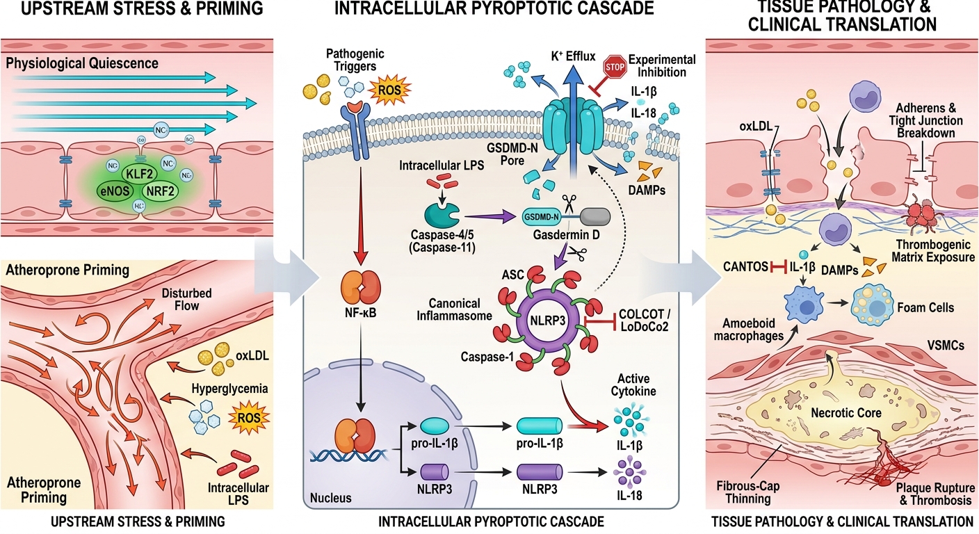
Graphical abstract
INTRODUCTION
Cardiovascular disease (CVD) remains the principal cause of morbidity and mortality globally, driven fundamentally by chronic, non-resolving vascular inflammation1. Within the arterial wall, chronic inflammatory signaling initiates endothelial dysfunction, compromises barrier integrity, augments endothelial permeability to circulating apolipoprotein B-containing lipoproteins, and drives sustained leukocyte recruitment and platelet activation1. Monocyte-derived macrophages within the subendothelial space exhibit phenotypic plasticity, adopting pro-inflammatory states that determine plaque fate—either lesion progression or resolution. Oxidized low-density lipoproteins (oxLDL) and triglyceride-rich remnant lipoproteins further ignite vascular inflammation by stimulating macrophages and vascular smooth muscle cells (VSMCs) to secrete potent pro-inflammatory cytokines, including interleukin-1β (IL-1β) and interleukin-6 (IL-6). These cytokines accelerate foam cell formation, expand necrotic lipid cores, and degrade extracellular matrix components within the protective fibrous cap. Persistent inflammatory signaling ultimately promotes vulnerable plaque development and catastrophic luminal thrombosis, cementing inflammation as a cornerstone pathogenic mechanism and a paramount therapeutic target in modern cardiovascular medicine1.
At the molecular level, inflammasome signaling complexes integrate noxious metabolic, oxidative, and hemodynamic stressors to orchestrate vascular and immune pathology. Both canonical and non-canonical inflammasome pathways operate within the vascular endothelium, governing the maturation and secretion of inflammatory cytokines and directing programmed cell death programs. Although the structural dynamics and activation kinetics of these complexes are delineated in subsequent sections, their specific role in precipitating endothelial barrier breakdown warrants early emphasis.
Pyroptosis has emerged as a distinct, highly pro-inflammatory form of regulated cell death (RCD) executed downstream of inflammasome assembly. Inflammatory caspase-mediated cleavage of the pore-forming effector gasdermin D (GSDMD) generates an N-terminal fragment (GSDMD-NT) that oligomerizes within the plasma membrane, forming transmembrane pores that trigger cell swelling, membrane rupture, and the massive release of IL-1β, interleukin-18 (IL-18), and damage-associated molecular patterns (DAMPs). Unlike apoptosis, which maintains plasma membrane integrity and is immunologically silent, pyroptosis is inherently immunogenic because it couples rapid cell lysis with the active extrusion of mature pyrogenic cytokines. Substantial preclinical and clinical evidence connects pyroptotic death pathways to acute myocardial infarction, myocarditis, chronic heart failure, atherosclerosis, arterial hypertension, and cardiac arrhythmias, underscoring its broad pathological footprint across the cardiovascular disease spectrum2,3.
In acute infectious settings, such as bacterial sepsis, controlled pyroptosis aids host defense by eliminating pathogen replication niches. However, hyperactivated inflammasome signaling triggers widespread endothelial damage, hyperpermeability, capillary leak syndrome, and multi-organ failure4. While sepsis illustrates the catastrophic potential of uncontrolled systemic pyroptosis, the present review focuses specifically on the localized and chronic mechanisms of endothelial pyroptosis within the cardiovascular continuum4.
The vascular endothelium is far more than a passive anatomical barrier; it operates as an active, highly sensitive endocrine and immune organ vital for hemodynamic and immunological homeostasis. Endothelial dysfunction represents the earliest detectable harbinger of atherogenesis and is intimately linked to chronic vascular inflammation, inflammasome assembly, and pyroptotic cell death2,3,4,5. Among the diverse inflammatory mediators involved, IL-1β occupies a central hierarchy by disrupting junctional complexes, inducing endothelial adhesion molecule expression, amplifying cytokine signaling cascades, and accelerating atherosclerotic plaque destabilization5. These cell-type-specific inflammatory pathways distinguish endothelial pyroptosis as a major driver of arterial disease and identify IL-1β-centric signaling as a compelling therapeutic candidate. Accordingly, this review examines the molecular machinery of endothelial pyroptosis, its mechanistic contribution to cardiovascular disease progression, and emerging translational avenues for therapeutic intervention4,5.
METHODOLOGY
This manuscript was formulated as a narrative review to provide an authoritative, comprehensive synthesis of current scientific evidence regarding IL-1β-mediated pyroptotic signaling in vascular endothelial cells and its role in cardiovascular disease progression. The review methodology was structured in accordance with the Scale for the Assessment of Narrative Review Articles (SANRA) guidelines to ensure methodological rigor, transparency, and comprehensive reporting quality.
A systematic literature search was executed across PubMed/MEDLINE, Scopus, Web of Science, and Google Scholar for peer-reviewed studies published between January 2010 and March 2025. The search architecture combined Medical Subject Headings (MeSH) and free-text terms using Boolean logic. Representative search terms included: ("pyroptosis" OR "inflammasome" OR "gasdermin") AND ("IL-1β" OR "Interleukin-1 beta" OR "caspase-1") AND ("endothelial cells" OR "vascular endothelium" OR "endothelial dysfunction") AND ("atherosclerosis" OR "cardiovascular disease" OR "vascular inflammation" OR "plaque vulnerability").
Additional eligible studies were identified through backward and forward citation tracking of included articles and landmark reviews. Original research articles, mechanistic in vitro and in vivo studies, randomized controlled clinical trials, systematic reviews, and meta-analyses investigating endothelial pyroptosis, inflammasome biology, IL-1β signaling, and cardiovascular outcomes were prioritized. Inclusion was restricted to English-language peer-reviewed publications. Articles unrelated to cardiovascular biology, studies focusing exclusively on non-vascular tissues without mechanistic extrapolation, conference abstracts lacking complete methodological details, and non-peer-reviewed commentaries were excluded. Retrieved publications were evaluated independently by title, abstract, and full-text screening, and synthesized into thematic sections addressing molecular mechanisms, cellular cross-talk, clinical trial evidence, and therapeutic perspectives.
VASCULAR ENDOTHELIUM IN CARDIOVASCULAR HOMEOSTASIS AND DISEASE
The vascular endothelium comprises a specialized, continuous monolayer of endothelial cells that lines the entire circulatory tree, serving as the interface between the bloodstream and underlying vascular tissues. Under physiological conditions, endothelial cells dynamically integrate mechanical forces (fluid shear stress) and biochemical cues to govern vascular tone, hemostatic balance, and inflammatory responses. They synthesize and release key vasoactive mediators, including vasodilators such as nitric oxide (NO) and prostacyclin (PGI₂), balanced against vasoconstrictors such as endothelin-1 (ET-1) and thromboxane A₂ (TXA₂). Furthermore, quiescent endothelial cells express cell-surface thrombomodulin and heparan sulfate proteoglycans, presenting an antithrombotic, anti-adhesive surface that inhibits platelet aggregation and restricts leukocyte transendothelial migration6,7.
Endothelial architecture, transcriptional programs, and physiological capabilities vary considerably across vascular beds, reflecting substantial endothelial heterogeneity. Cerebral microvascular endothelial cells assemble continuous, high-resistance tight junctions that form the blood–brain barrier, whereas renal glomerular and hepatic sinusoidal endothelial cells possess fenestrations and discontinuities tailored for filtration and macromolecular exchange7. Beyond cross-organ differences, arterial endothelial cells residing within coronary, carotid, aortic, and peripheral conduit vessels manifest distinct phenotypic responses to local hemodynamics8. Under steady, laminar blood flow, high shear stress maintains endothelial cells in a quiescent, anti-inflammatory, and antithrombotic state. This mechanoprotection is orchestrated via Krüppel-like factor 2 (KLF2), endothelial nitric oxide synthase (eNOS) activation, and nuclear factor erythroid 2-related factor 2 (NRF2)-mediated antioxidant gene expression. Conversely, disturbed or oscillatory shear stress—predominant at arterial bifurcations, branch points, and outer curvatures—suppresses KLF2 and eNOS while activating sterol regulatory element-binding proteins (SREBPs) and nuclear factor kappa B (NF-κB). This biomechanical stress elevates reactive oxygen species (ROS) production, primes the NLRP3 inflammasome, and renders atheroprone endothelium uniquely susceptible to pyroptotic cell death6,8. These regional hemodynamic discrepancies explain the focal predilection of atherosclerotic plaques for specific anatomical sites along the arterial tree.
Endothelial dysfunction, characterized by diminished NO bioavailability, profound oxidative stress, and a phenotypic shift toward a pro-inflammatory, pro-thrombotic state, constitutes an indispensable early event in cardiovascular pathogenesis. Conventional cardiovascular risk factors—including essential hypertension, diabetes mellitus, dyslipidemia, cigarette smoking, and biological aging—impair endothelium-dependent vasodilation and compromise junctional integrity. Together, these systemic insults establish a permissive vascular microenvironment that fosters sustained vascular inflammation, inflammasome priming, and endothelial pyroptosis, directly fueling disease initiation and progression6,9,10.
Inflammasome signaling serves as a critical biochemical transducing hub within this dysfunctional milieu. Upon exposure to cellular stress, endothelial cells activate canonical and non-canonical inflammasome cascades, resulting in caspase activation, inflammatory cytokine processing, and GSDMD cleavage11. This inflammatory response is self-amplifying: pro-inflammatory mediators including tumor necrosis factor-alpha (TNF-α), IL-6, IL-1β, and oxidized phospholipids induce the expression of vascular cell adhesion molecule 1 (VCAM-1), intercellular adhesion molecule 1 (ICAM-1), and E-selectin, promoting leukocyte docking and diapedesis. Concomitant activation of canonical NF-κB pathways enhances oxidative stress through NADPH oxidases (NOX2 and NOX4), which uncouple eNOS and exhaust NO reserves. Synergistic interactions involving angiotensin II, CD40/CD40 ligand signaling, and transforming growth factor-beta (TGF-β) further sustain this pro-inflammatory, pro-fibrotic state, accelerating vascular remodeling and atherosclerosis12,13.
Importantly, endothelial pyroptosis must be distinguished mechanistically and functionally from macrophage pyroptosis. While macrophage pyroptosis represents an innate immune defense response that expands plaque necrotic cores upon excessive lipid loading2, endothelial pyroptosis directly breaches the vascular barrier6,13. Endothelial cell lysis strips the luminal lining, exposes the highly thrombogenic subendothelial basement membrane, provokes microvascular thrombosis, accelerates vascular leak, and grants circulating leukocytes and atherogenic lipoproteins direct access to the intima6,12,13. Consequently, the pathological repercussions of endothelial pyroptosis extend far beyond cytokine release, directly destabilizing vascular architecture and vascular homeostasis2,6. Given that this vulnerability is strongly modulated by local shear stress and arterial bed identity, endothelial pyroptosis operates as a regionally tuned driver of cardiovascular disease8,13.
In acute systemic conditions such as sepsis, endothelial pyroptosis underlies microvascular leakage, capillary thrombosis, and refractory distributive shock. Controlled pyroptosis provides antibacterial host defense, but widespread, unconstrained endothelial pore formation precipitates diffuse vascular collapse and multi-organ dysfunction4.
PYROPTOSIS: AN INFLAMMATORY MODE OF PROGRAMMED CELL DEATH
Pyroptosis is a distinct form of regulated cell death (RCD) driven by inflammatory caspases and executed by pore-forming gasdermin proteins. Originally described in macrophages resisting intracellular pathogens, pyroptosis is now established as a ubiquitous stress-response pathway operating in parenchymal, immune, and vascular tissues. While apoptosis preserves plasma membrane integrity and results in apoptotic body phagocytosis without inflammatory collateral damage, pyroptosis is intrinsically lytic and fiercely pro-inflammatory, culminating in rapid plasma membrane rupture and the extracellular discharge of cytoplasmic contents and mature cytokines. Mechanistically, inflammatory caspases cleave gasdermin D (GSDMD), liberating its cytotoxic N-terminal domain (GSDMD-NT). GSDMD-NT fragments bind specifically to phosphoinositides on the inner leaflet of the plasma membrane and oligomerize into large transmembrane pores comprising 24–33 protomers with an internal diameter of 10–15 nm. These macroscopic pores disrupt transmembrane ionic gradients, permitting unrestricted potassium efflux accompanied by sodium and water influx. The resulting oncotic pressure drives rapid cellular swelling, ballooning, and catastrophic membrane breakdown, while simultaneously providing a conduit for the unconventional secretion of leaderless cytokines (IL-1β and IL-18) and potent DAMPs14.
Morphologically and biochemically, pyroptosis is distinguished by early membrane permeabilization, rapid propidium iodide/ethidium bromide uptake, chromatin condensation without karyorrhexis or internucleosomal DNA laddering, and massive release of cytosolic enzymes such as lactate dehydrogenase (LDH) and high mobility group box 1 (HMGB1), which profoundly amplify regional and systemic inflammation15.
Although apoptosis, pyroptosis, and necroptosis are categorized as major regulated cell death modalities, they exhibit distinct molecular triggers, signaling cascades, and immunological outcomes (Table 1). Apoptosis is orchestrated by initiator caspases (caspase-8, -9) and executioner caspases (caspase-3, -7), resulting in cell shrinkage, membrane blebbing, DNA internucleosomal fragmentation, and formation of intact apoptotic bodies cleared by efferocytosis without immune provocation. In contrast, pyroptosis is executed via caspase-1, -4, -5, or -11 and GSDMD pore formation, driving explosive inflammatory cytokine secretion. Necroptosis represents an alternative inflammatory cell death program that is caspase-independent and regulated by the receptor-interacting serine/threonine-protein kinase 1 (RIPK1), RIPK3, and mixed lineage kinase domain-like protein (MLKL) axis, typically initiated upon death receptor ligation under caspase-8-inhibited conditions. Although necroptosis also causes cell lysis and DAMP release, it occurs independently of inflammasome assembly and gasdermin cleavage. Notably, intricate cross-talk unites these death pathways; for example, apoptotic caspase-3 can cleave gasdermin E (GSDME) to induce secondary pyroptotic lysis, and components of the PANoptosome complex can co-regulate apoptosis, necroptosis, and pyroptosis in response to complex microbial or metabolic insults16,17.
Distinguishing biochemical, morphological, and immunological characteristics of programmed cell death modalities in vascular pathophysiology
| Feature | Apoptosis | Pyroptosis | Necroptosis |
|---|---|---|---|
| Primary Triggers | Intrinsic mitochondrial pathway (DNA damage, oxidative stress, developmental cues) or extrinsic death receptor signaling (FasL, TRAIL). | Pathogen- and danger-associated molecular patterns (PAMPs/DAMPs, e.g., oxLDL, cholesterol crystals, disturbed shear stress, cytosolic LPS, extracellular ATP). | Death receptor ligation (TNF-α, FasL, TRAIL) in the presence of caspase-8 inhibition or depletion. |
| Key Molecular Effectors | Initiator caspases (caspase-8, -9); executioner caspases (caspase-3, -7); Bcl-2 family proteins. | Inflammatory caspases: caspase-1 (canonical); caspase-4/5 in humans and caspase-11 in mice (non-canonical); pore-forming executioner Gasdermin D (GSDMD). | Receptor-interacting serine/threonine-protein kinases 1 and 3 (RIPK1, RIPK3); mixed lineage kinase domain-like protein (MLKL). |
| Membrane Integrity & Kinetics | Maintained throughout early and intermediate phases; preservation of membrane impermeability with exposure of external phosphatidylserine; late-stage apoptotic bodies. | Rapidly permeabilized via GSDMD-NT oligomeric pores (10–15 nm); early loss of electrochemical gradients, cellular oncotic swelling, and explosive lysis. | Disrupted via MLKL oligomerization and plasma membrane translocation; cell swelling and necrotic plasma membrane rupture. |
| Tissue Inflammatory Response | Immunologically silent or anti-inflammatory; cellular fragments contained in apoptotic bodies efficiently cleared by scavenger macrophage efferocytosis. | Fiercely pro-inflammatory; massive release of intracellular contents, pyrogenic cytokines, and alarmins into surrounding vascular tissue. | Pro-inflammatory; immunogenic lytic release of intracellular DAMPs into the vascular microenvironment. |
| Cytokine & Mediator Release | No active cytokine maturation or lytic release; minimal inflammatory alarmin discharge. | Active maturation and massive unconventional release of IL-1β and IL-18 via GSDMD pores; passive extrusion of DAMPs (HMGB1, ATP, S100 proteins). | No active IL-1β/IL-18 cytokine maturation; extensive passive release of DAMPs (HMGB1, mitochondrial DNA, IL-1α, IL-33). |
Within the vascular wall, pyroptosis carries unique pathological gravity. Endothelial cell death directly dismantles the protective vascular barrier, elevates permeability, accelerates plasma exudation, exposes thrombogenic collagen, and facilitates extensive leukocyte infiltration6,7,13. Furthermore, pyroptotic endothelial cells release mediator clouds that engage surrounding VSMCs and macrophages, fueling a chronic inflammatory spiral that propels atheroma growth and destabilization2,6,13,17. Hence, endothelial pyroptosis operates not merely as an end-stage marker of cell injury, but as an active, disease-amplifying engine in cardiovascular pathology2,6,17.
Both canonical and non-canonical inflammasome pathways converge on GSDMD pore formation, despite deploying distinct upstream sensors and initiating caspases18. Their precise molecular architecture and endothelial contributions are dissected below.
MOLECULAR MACHINERY OF PYROPTOSIS
The execution of pyroptosis requires multiprotein inflammasome assembly, inflammatory caspase activation, and gasdermin-mediated membrane perforation. These complexes sense pathogen-associated molecular patterns (PAMPs) and danger-associated molecular patterns (DAMPs) to coordinate cellular defense or pathological tissue destruction18. Canonical inflammasomes consist of a cytosolic pattern-recognition receptor (PRR) sensor (e.g., NLRP3, AIM2, or NLRC4), the bipartite adaptor protein ASC (apoptosis-associated speck-like protein containing a CARD), and the zymogen pro-caspase-1. Sensing of intracellular disturbances initiates sensor oligomerization, which recruits ASC via pyrin domain (PYD) interactions. ASC molecules then assemble into large macromolecular "specks" that present their caspase activation and recruitment domains (CARD) to recruit pro-caspase-1, triggering proximity-induced autocatalytic cleavage. Active caspase-1 subsequently processes inactive pro-IL-1β and pro-IL-18 into mature cytokines while simultaneously cleaving GSDMD, coupling cytokine maturation directly to cell lysis19.
Among canonical sensors, NLRP3 (NOD-like receptor family pyrin domain-containing 3) is uniquely prominent in vascular biology because it integrates an exceptionally diverse array of non-microbial metabolic and hemodynamic stressors. NLRP3 activation is initiated by potassium (K⁺) efflux, mitochondrial ROS generation, mitochondrial DNA translocation, cardiolipin exposure, lysosomal rupture with cathepsin B release, and calcium flux, with the mitotic kinase NEK7 serving as an indispensable catalytic licensing binding partner19. In contrast, AIM2 (absent in melanoma 2) specifically binds cytosolic double-stranded DNA via its HIN-200 domain, while NLRC4 detects intracellular bacterial flagellin and type III secretion apparatus rod components via NLR family apoptosis inhibitory proteins (NAIPs)20.
In endothelial cells, the contribution of individual inflammasome complexes is governed by the prevailing microenvironmental milieu. Pro-atherogenic insults—including oscillatory shear stress, cholesterol crystals, oxLDL, high ambient glucose, and mitochondrial dysfunction—activate the endothelial NLRP3 inflammasome, accelerating pyroptotic injury6,13,18. This context-dependent activation is critical, as endothelial cells from distinct vascular beds exhibit marked heterogeneity in their metabolic and inflammatory thresholds13.
The canonical pathway requires a two-step signaling mechanism (Figure 1). Priming (Signal 1) is initiated by ligands binding to TLRs, IL-1R1, or TNF receptors, stimulating NF-κB-dependent transcription of NLRP3, pro-IL-1β, and pro-IL-18, alongside post-translational deubiquitination of NLRP3. Activation (Signal 2) is triggered by metabolic or physical disturbances (e.g., K⁺ efflux, ROS), culminating in ASC speck formation, caspase-1 activation, cytokine cleavage, and GSDMD-mediated membrane permeabilization18,19.
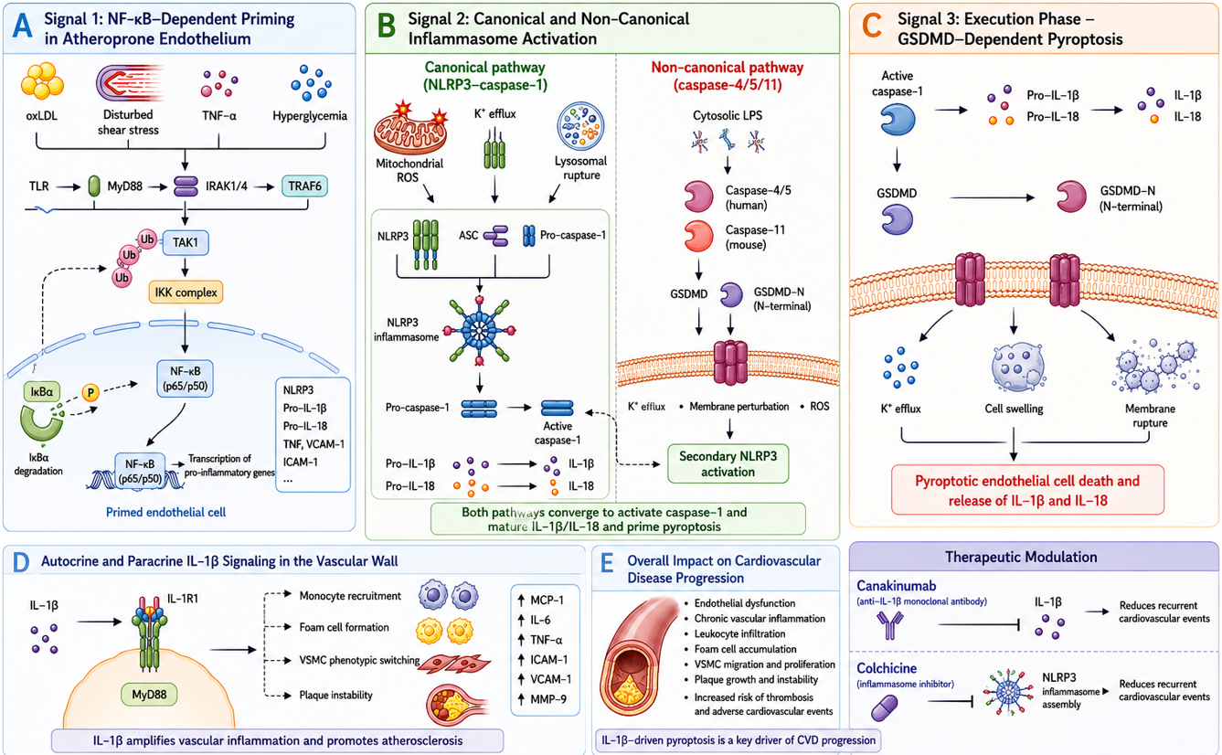
Molecular mechanisms of IL-1β-centric canonical and non-canonical inflammasome signaling in endothelial pyroptosis, vascular remodeling, and cardiovascular disease progression. Schematic illustration delineating the multistep priming, activation, execution, and intercellular propagation of interleukin-1β (IL-1β)-driven endothelial pyroptosis within the arterial wall, alongside therapeutic points of intervention evaluated in clinical trials. Panel A | Signal 1: NF-κB–Dependent Priming in Atheroprone Endothelium. Pro-atherogenic cardiovascular stressors—including oxidized low-density lipoprotein (oxLDL), disturbed or oscillatory shear stress, tumor necrosis factor-alpha (TNF-α), and hyperglycemia—engage Toll-like receptors (TLRs) and cytokine receptors on the endothelial luminal surface. Downstream recruitment of myeloid differentiation primary response 88 (MyD88), interleukin-1 receptor-associated kinases 1 and 4 (IRAK1/4), and TNF receptor-associated factor 6 (TRAF6) activates transforming growth factor-β-activated kinase 1 (TAK1) and the IκB kinase (IKK) complex. IKK induces phosphorylation, polyubiquitination (Ub), and proteasomal degradation of the inhibitory chaperone IκBα, enabling liberated NF-κB heterodimers (p65/p50) to translocate into the nucleus. This transcriptional priming drives the upregulation of core inflammasome components and pro-inflammatory mediators, including NLRP3, pro-IL-1β, pro-IL-18, TNF-α, vascular cell adhesion molecule 1 (VCAM-1), and intercellular adhesion molecule 1 (ICAM-1), in primed endothelial cells. Panel B | Signal 2: Canonical and Non-Canonical Inflammasome Activation. Canonical inflammasome assembly is triggered when primed endothelial cells encounter secondary danger signals, including mitochondrial reactive oxygen species (ROS), intracellular potassium (K⁺) efflux, and lysosomal membrane destabilization. These signals induce the sensor NLRP3 to oligomerize and recruit apoptosis-associated speck-like protein containing a CARD (ASC) and pro-caspase-1, forming the multimeric NLRP3 inflammasome complex. Proximity-induced autocatalytic activation generates active caspase-1, which proteolytically cleaves inactive 31-kDa pro-IL-1β and pro-IL-18 into mature, bioactive 17-kDa IL-1β and IL-18. In parallel, the non-canonical pathway is initiated by direct intracellular sensing of bacterial lipopolysaccharide (LPS) by human caspase-4 and caspase-5 (or murine caspase-11), which directly cleaves gasdermin D (GSDMD). GSDMD-mediated membrane permeabilization promotes subsequent K⁺ efflux and cellular stress, triggering secondary activation of the canonical NLRP3–caspase-1 axis to ensure cytokine processing. Panel C | Signal 3: Execution Phase – GSDMD-Dependent Pyroptosis. Active caspase-1 (and non-canonical caspases-4/5/11) proteolytically cleaves full-length GSDMD (53 kDa), releasing its autoinhibitory C-terminal domain and liberating the cytotoxic N-terminal fragment (GSDMD-N, 31 kDa). GSDMD-N monomers oligomerize within the inner leaflet of the endothelial plasma membrane, forming symmetrical transmembrane channels (10–15 nm inner diameter). Uncontrolled pore opening collapses transmembrane electrochemical gradients, driving rapid K⁺ efflux, sodium and water influx, cellular swelling, ballooning, and catastrophic plasma membrane rupture (pyroptosis). This lytic event facilitates the massive, passive unconventional extrusion of mature IL-1β, IL-18, and intracellular damage-associated molecular patterns (DAMPs) into the subendothelial space. Panel D | Autocrine and Paracrine IL-1β Signaling in the Vascular Wall. Released extracellular IL-1β engages interleukin-1 receptor type 1 (IL-1R1) on endothelial cells, adjacent vascular smooth muscle cells (VSMCs), and infiltrating monocytes/macrophages. Signal transduction through MyD88 stimulates robust downstream secretion of monocyte chemoattractant protein-1 (MCP-1), interleukin-6 (IL-6), TNF-α, and matrix metalloproteinase-9 (MMP-9), alongside pronounced cell-surface upregulation of VCAM-1 and ICAM-1. This establishes an autocrine and paracrine feed-forward loop that accelerates monocyte recruitment, macrophage foam cell formation, phenotypic switching of VSMCs from a quiescent contractile to a proliferative synthetic state, and extracellular matrix degradation. Panel E | Overall Impact on Cardiovascular Disease Progression and Therapeutic Modulation. The cumulative biological effects of endothelial pyroptosis culminate in vascular barrier disruption, chronic vascular inflammation, dense leukocyte infiltration, foam cell accumulation, necrotic core enlargement, fibrous cap thinning, and heightened susceptibility to plaque rupture and luminal thrombosis. Pharmacological interventions targeting this axis have demonstrated clinical efficacy in reducing major adverse cardiovascular events (MACE): canakinumab (a human monoclonal antibody selectively neutralizing IL-1β) demonstrated significant cardiovascular risk reduction in the CANTOS trial, while low-dose colchicine (which inhibits microtubule dynamics and NLRP3 inflammasome assembly) significantly reduced ischemic events in the COLCOT and LoDoCo2 trials.
Non-canonical pyroptosis operates through distinct caspases depending on the mammalian species. In humans, intracellular lipopolysaccharide (LPS) from Gram-negative bacteria directly engages the CARD domains of caspase-4 and caspase-5, whereas in mice, this direct recognition is mediated by caspase-1121,22. LPS binding triggers oligomerization and activation of caspase-4/5/11 without requiring classical PRR sensors or ASC adaptors. Active caspase-4/5/11 efficiently cleaves GSDMD, generating GSDMD-NT and inducing lytic membrane perforation. Although these non-canonical caspases cannot directly cleave pro-IL-1β or pro-IL-18 efficiently, the resulting GSDMD pores induce robust K⁺ efflux, which secondarily triggers canonical NLRP3 inflammasome assembly, thereby enabling caspase-1-dependent cytokine processing and secretion23,24.
Thus, canonical and non-canonical signaling pathways converge on GSDMD cleavage, with the non-canonical pathway engaging secondary NLRP3 activation to achieve complete cytokine maturation. While non-canonical mechanisms have been studied predominantly in immune cells, their contribution to endothelial damage in endotoxemia and vascular inflammation is increasingly recognized and must be interpreted in light of human–murine caspase divergences21,23,24.
Gasdermins represent the terminal pore-forming executioners of pyroptosis. Full-length GSDMD (53 kDa) is held in an autoinhibited state through intramolecular interaction between its C-terminal repressor domain (GSDMD-CT) and its N-terminal pore-forming domain (GSDMD-NT). Cleavage at Asp275 (human) or Asp276 (mouse) relieves this autoinhibition, allowing the 31-kDa GSDMD-NT to oligomerize and form nonselective transmembrane channels21. Uncontrolled pore formation dissipates cellular ionic gradients, drives cell swelling, and precipitates membrane rupture. Concurrently, the endosomal sorting complexes required for transport (ESCRT) machinery, particularly ESCRT-III, can be recruited to the plasma membrane to shed pore-bearing membrane vesicles, transiently opposing cell death when the pyroptotic stimulus is submaximal25.
Within the vascular endothelium, pyroptotic pore formation compromises barrier integrity and accelerates inflammatory interactions with circulating and resident vascular cells13,18. These molecular lesions promote endothelial detachment, enhance leukocyte transmigration, and amplify communication between endothelial cells, macrophages, and VSMCs, firmly integrating pyroptotic execution with chronic vascular inflammation6,13,18,19 (Figure 1).
IL-1β BIOGENESIS AND SIGNALING PATHWAYS
The biogenesis, maturation, and signaling cascades of interleukin-1β (IL-1β) are under stringent multilayered regulation to prevent uncontrolled inflammatory damage in response to sterile injury, infection, or metabolic stress. In quiescent endothelial cells and leukocytes, basal IL1B transcription is negligible. Induction requires an initial priming stimulus (Signal 1), typically delivered through pattern-recognition receptors (e.g., TLR2, TLR4) or cytokine receptors (e.g., IL-1R1, TNFR1). Downstream activation of NF-κB and mitogen-activated protein kinases (MAPKs) directs the binding of transcription factors—including NF-κB p65/p50, activator protein-1 (AP-1; c-Jun/c-Fos), CCAAT/enhancer-binding protein beta (C/EBPβ), and interferon regulatory factors (IRFs)—to the IL1B promoter, driving the synthesis of a 31-kDa inactive zymogen, pro-IL-1β26. Cell-type-specific transcriptional networks fine-tune this response; for instance, the pioneer factor Spi-1/PU.1 coordinates with C/EBPβ in myeloid lineages, whereas endothelial cells rely heavily on NF-κB and AP-1 complexes. Furthermore, chromatin accessibility is modulated by histone post-translational modifications (H3K4me3 and H3K27me3), while metabolic intermediates such as succinate and fumarate accumulate during metabolic reprogramming to stabilize hypoxia-inducible factor 1-alpha (HIF-1α), sustaining sustained IL1B transcription27.
Unlike classical leader-containing secretory proteins that traverse the endoplasmic reticulum–Golgi pathway, pro-IL-1β is synthesized in the cytosol without an N-terminal signal peptide and remains biologically inert. To acquire biological potency, pro-IL-1β must undergo proteolytic processing. Caspase-1, activated within canonical inflammasomes, cleaves pro-IL-1β at the specific peptide bond Asp116–Ala117 (in human pro-IL-1β), liberating the 17-kDa mature, fully active cytokine28.
Following proteolytic cleavage, mature IL-1β is released into the extracellular space. GSDMD-dependent plasma membrane pores represent the primary exit route during pyroptosis28. However, in non-pyroptotic contexts or during hyperactivation states, alternative mechanisms—including purinergic P2X7 receptor-dependent microvesicle shedding, multivesicular body exocytosis, and chaperone-mediated autophagy pathways—can facilitate unconventional IL-1β secretion independently of lytic cell death29.
Non-canonical inflammasome pathways also contribute indirectly to IL-1β processing. Human caspase-4/5 and murine caspase-11 do not cleave pro-IL-1β directly; instead, their cleavage of GSDMD and subsequent pore-mediated K⁺ efflux triggers secondary assembly of the canonical NLRP3 inflammasome, which supplies active caspase-1 for pro-IL-1β maturation16. In specialized settings, alternative proteases—such as caspase-8 in TLR-primed apoptotic platforms, neutrophil elastase, proteinase 3, cathepsin G, and mast cell chymase—can cleave pro-IL-1β into bioactive forms, providing caspase-1-independent pathways of cytokine activation in inflamed tissues16.
Extracellular mature IL-1β binds with high affinity to its cognate surface receptor, interleukin-1 receptor type 1 (IL-1R1), inducing the recruitment of the interleukin-1 receptor accessory protein (IL-1RAcP) coreceptor to form a high-affinity signaling complex. The intracellular Toll/IL-1 receptor (TIR) domains of IL-1R1 and IL-1RAcP recruit the adaptor protein myeloid differentiation primary response 88 (MyD88), which nucleates the "myddosome" complex with interleukin-1 receptor-associated kinases (IRAK1, IRAK4). Phosphorylated IRAKs dissociate and engage TNF receptor-associated factor 6 (TRAF6), triggering Lys63-linked polyubiquitination that recruits and activates transforming growth factor-β-activated kinase 1 (TAK1). Activated TAK1 simultaneously stimulates the IκB kinase (IKK) complex (leading to IκBα degradation and NF-κB nuclear translocation) and MAPK signaling pathways (p38, JNK, ERK1/2)30. This robust transcriptional cascade drives the expression of secondary inflammatory cytokines (IL-6, TNF-α), chemokines (MCP-1/CCL2, IL-8/CXCL8), matrix metalloproteinases (MMP-2, MMP-9), and vascular adhesion molecules (VCAM-1, ICAM-1, E-selectin), fueling local tissue inflammation and remodeling.
Under physiological conditions, IL-1β signaling is constrained by endogenous regulatory checkpoints, including the competitive decoy receptor IL-1R2, the soluble antagonist interleukin-1 receptor antagonist (IL-1Ra), and oxygen-sensing prolyl hydroxylases31. However, sustained metabolic, oxidative, and hemodynamic stress disrupts these inhibitory mechanisms, producing unchecked IL-1β production, persistent vascular endothelial activation, and progressive cardiovascular pathology32.
IL-1β-CENTRIC PYROPTOTIC SIGNALING IN VASCULAR ENDOTHELIUM
Vascular endothelial cells, traditionally considered passive physical conduits, are now recognized as competent, non-classical innate immune sentinels equipped to mount robust inflammasome-driven responses. In cardiovascular disease, endothelial cells are continuously exposed to metabolic, oxidative, and biomechanical stressors, including oxidized low-density lipoprotein (oxLDL), disturbed shear stress, hyperglycemia, cholesterol crystals, and mitochondrial reactive oxygen species (ROS). These pathological stimuli induce endothelial activation and elevate susceptibility to inflammasome-associated injury33.
Under sustained or severe cellular stress, endothelial NLRP3 inflammasome activity increases, promoting inflammatory cytokine maturation and pyroptotic execution18,19,33. Rather than serving merely as an isolated cell death pathway, endothelial pyroptosis fundamentally alters the local vascular microenvironment by coupling structural cellular disruption with the burst release of pro-inflammatory mediators33.
Preclinical studies demonstrate that endothelial NLRP3 activation directly increases IL-1β secretion and precipitates pyroptotic endothelial damage33. This signaling cascade establishes an inflammatory vascular niche, linking metabolic and biomechanical stress to sustained endothelial dysfunction and cardiovascular disease progression33.
Pyroptotic injury severely compromises endothelial barrier integrity and disrupts homeostatic interactions between the endothelium, circulating leukocytes, and resident vascular cells13,33. GSDMD pore formation and cell lysis loosen intercellular junctions, increase permeability to circulating lipoproteins, and expose the thrombogenic subendothelial matrix. These structural alterations stimulate leukocyte adhesion and transmigration and heighten inflammatory signaling within the arterial wall18,33. Thus, endothelial pyroptosis propagates vascular inflammation well beyond the initially injured cells13,18,33.
Non-canonical inflammasome pathways also participate in endothelial inflammatory damage, particularly via caspase-4 and caspase-5 in human endothelial cells21,23,24. Although preclinical studies support their contribution to endothelial dysfunction, much of the foundational mechanistic insight derives from myeloid models. Consequently, the precise clinical contribution of non-canonical pyroptosis to human atherogenesis remains less fully defined than that of the canonical NLRP3–caspase-1–IL-1β axis.
Extracellularly released IL-1β exerts powerful autocrine and paracrine actions across the vascular wall. In an autocrine loop, IL-1β binds endothelial IL-1R1 to reactivate NF-κB, upregulating adhesion molecules (VCAM-1, ICAM-1) and chemokines (MCP-1), which accelerates leukocyte recruitment. In paracrine circuits, endothelial-derived IL-1β stimulates neighboring VSMCs and infiltrating macrophages, promoting cytokine release and matrix degradation. Supporting this pathogenic paradigm, studies in apolipoprotein E-deficient (ApoE) mice demonstrate that genetic deletion or pharmacological inhibition of IL-1β significantly suppresses vascular inflammation and attenuates atherosclerotic lesion development34,6.
In endothelial cells, IL-1β signaling reinforces pro-inflammatory transcriptional programs and amplifies ROS production, establishing a feed-forward inflammatory circuit that maintains chronic endothelial activation35.
Collectively, these data substantiate an IL-1β-centered feed-forward model: cardiovascular stressors prime and activate endothelial inflammasomes, inducing pyroptotic injury and cytokine release, while extracellular IL-1β reinforces NF-κB-dependent inflammation within adjacent vascular and immune cells35,36. This reciprocal loop between endothelial damage and vascular inflammation sustains the progression of cardiovascular disease.
ROLE OF ENDOTHELIAL PYROPTOSIS IN CARDIOVASCULAR DISEASE PROGRESSION
Atherosclerosis represents the primary pathological substrate of cardiovascular disease, evolving from intimal lipid retention to the formation of structurally divergent plaques that dictate distinct clinical outcomes. Crucially, plaque biological composition and vulnerability, rather than luminal stenosis alone, represent the primary determinants of acute cardiovascular events. High-risk, vulnerable plaques feature large necrotic lipid cores, thin fibrous caps (<65 μm), and dense inflammatory cell infiltration. These unstable features confer marked susceptibility to plaque rupture or surface erosion, triggering acute luminal thrombosis that manifests clinically as acute coronary syndromes (ACS), acute myocardial infarction, or sudden cardiac death. In contrast, stable plaques possess thick, collagen-rich fibrous caps and calcified matrices, typically manifesting as chronic stable angina or chronic total occlusions. Delineating plaques across this stability continuum clarifies the transition from indolent lesion formation to catastrophic vascular thrombosis37.
Endothelial pyroptosis directly contributes to atherosclerotic lesion initiation, plaque expansion, and fibrous cap destabilization. In early atherogenesis, endothelial cells exposed to disturbed shear forces, oxLDL, hypercholesterolemia, and metabolic stress assemble the NLRP3 inflammasome, driving caspase-1-dependent GSDMD cleavage and pro-IL-1β/pro-IL-18 maturation. Cleaved GSDMD-NT integrates into the plasma membrane to form cytolytic pores, collapsing ionic balance and causing lytic cell death. The resulting endothelial injury is accompanied by elevated ROS generation and sustained NF-κB signaling, promoting monocyte attachment, subendothelial migration, foam cell differentiation, and fatty streak expansion38.
As atherosclerotic plaques advance, persistent endothelial pyroptosis maintains high local cytokine levels, intensifies intraplaque inflammation, and weakens the protective fibrous cap by augmenting macrophage pyroptosis and enlarging the central necrotic core. Heightened endothelial IL-1β expression strongly correlates with histological features of plaque instability and elevated rupture risk33,39.
A central pathophysiological consequence of endothelial pyroptosis is the disruption of vascular barrier integrity. GSDMD pore formation and subsequent cell lysis disassemble tight junctions (claudin-5, occludin, zonula occludens-1) and adherens junctions (vascular endothelial-cadherin), inducing vascular hyperpermeability and interstitial edema. Both canonical and non-canonical pyroptotic cascades contribute to endothelial barrier breakdown. Through GSDMD-mediated membrane permeabilization and subsequent cytokine release, these pathways amplify vascular inflammation, permit excessive LDL influx, and accelerate atheroma development40.
Simultaneously, IL-1β signaling strongly upregulates endothelial adhesion molecules (VCAM-1, ICAM-1, E-selectin), accelerating leukocyte rolling, firm adhesion, and transendothelial extravasation. Chemokines released during pyroptotic lysis (e.g., MCP-1, IL-8) amplify the recruitment of monocytes and neutrophils, perpetuating vascular inflammation and foam cell accumulation38,41. Downstream activation of NF-κB and MAPK pathways further primes the inflammasome, establishing a self-amplifying feed-forward circuit that drives continuous immune cell infiltration42.
Beyond plaque progression, endothelial pyroptosis plays an active role in arterial thrombosis, acute ischemic injury, and maladaptive vascular remodeling. Pyroptotic endothelial cells shed their physiological antithrombotic surface, downregulating thrombomodulin and tissue factor pathway inhibitor (TFPI) while exposing procoagulant phosphatidylserine and releasing tissue factor (TF)-enriched extracellular microparticles that initiate extrinsic coagulation. Concomitant pyroptosis of plaque macrophages and VSMCs destabilizes the plaque shoulder, predisposing to fibrous cap rupture and occlusive thrombus formation43. In ischemia–reperfusion injury, endothelial pyroptosis exacerbates microvascular no-reflow, extravasation, and inflammatory cytokine storms, deepening myocardial hypoxia and hindering tissue repair43. Furthermore, chronic pyroptotic signaling promotes maladaptive vascular remodeling by driving VSMC phenotypic switching, matrix metalloproteinase-mediated matrix degradation, and adventitial fibrosis, which contribute to arterial stiffening and luminal narrowing in advanced vascular disease43.
The clinical relevance of endothelial pyroptosis is highlighted by clinical trials demonstrating that targeted inhibition of IL-1β signaling reduces recurrent major adverse cardiovascular events (MACE). These landmark findings confirm that IL-1β-driven vascular inflammation and endothelial pyroptosis represent central pathogenic drivers of cardiovascular disease progression and valid therapeutic targets44.
CROSS-TALK BETWEEN IL-1β-DRIVEN PYROPTOSIS AND OTHER CELL TYPES
IL-1β-mediated endothelial pyroptosis does not occur in isolation; it develops through dynamic reciprocal interactions with neighboring resident vascular cells and infiltrating leukocytes—most prominently macrophages and vascular smooth muscle cells (VSMCs)—generating a self-perpetuating inflammatory microenvironment that propels chronic cardiovascular disease progression45,46. Within atherosclerotic plaques, monocyte-derived macrophages are primary contributors to inflammasome activation and IL-1β production. Endothelial-derived IL-1β accelerates monocyte adhesion, transendothelial migration, and macrophage differentiation, whereas macrophage-derived IL-1β binds endothelial IL-1R1 to amplify endothelial inflammasome activation, forging a bidirectional inflammatory feedback loop that perpetuates vascular injury42. Moreover, pyroptotic macrophages release extracellular ASC specks and intact inflammasome oligomers into the extracellular space. These particulate aggregates are internalized by adjacent endothelial cells and VSMCs, triggering secondary inflammasome assembly and escalating local tissue damage45,46.
VSMCs are profoundly affected by IL-1β-dependent intercellular signaling. Exposure to IL-1β induces phenotypic switching of VSMCs from a quiescent, contractile state (characterized by smooth muscle α-actin and calponin expression) to a synthetic, pro-inflammatory phenotype marked by heightened cytokine secretion, enhanced migration, accelerated proliferation, and altered extracellular matrix turnover. Synthetic VSMCs downregulate collagen synthesis and upregulate matrix-degrading enzymes (MMP-2, MMP-9), weakening the fibrous cap and predisposing plaques to rupture47. Furthermore, chronic IL-1β exposure activates the NLRP3 inflammasome within VSMCs, inducing VSMC pyroptosis, vessel wall thinning, and dystrophic calcification that destabilize advanced atheromas48.
Persistent vascular inflammation is sustained through reciprocal signaling between immune cells and the endothelium, with IL-1β acting as the core molecular mediator. Endothelial IL-1β signaling upregulates adhesion molecules (VCAM-1, ICAM-1) and chemokines (MCP-1), sustaining continuous leukocyte infiltration across the vascular barrier. Recruited monocytes, macrophages, and neutrophils secrete additional IL-1β, TNF-α, and ROS, which re-prime endothelial inflammasomes through NF-κB-dependent mechanisms. This reciprocal feedback loop lowers the activation threshold for endothelial pyroptosis, maintains chronic endothelial activation, and sustains immune recruitment, transforming transient inflammatory stimuli into non-resolving vascular inflammation characteristic of progressive atherosclerosis and related cardiovascular disorders43,49.
Although endothelial pyroptosis initiates locally within the vessel wall, its consequences reverberate systemically, contributing to generalized inflammatory activation and elevated cardiovascular risk. IL-1β released from pyroptotic endothelial cells enters the systemic circulation and acts on the liver to stimulate the hepatic acute-phase response (elevating high-sensitivity C-reactive protein [hsCRP] and fibrinogen), promotes bone marrow leukopoiesis, activates circulating leukocytes, and induces systemic endothelial dysfunction. Systemic inflammasome activation is a hallmark of cardiometabolic comorbidities, including type 2 diabetes mellitus, obesity, chronic kidney disease, and arterial hypertension, positioning endothelial pyroptosis as a functional nexus connecting localized arterial damage to systemic inflammatory burden44,49. In severe inflammatory conditions such as sepsis and severe viral infections, widespread endothelial pyroptosis causes capillary leak syndrome, disseminated intravascular coagulation (DIC), and inflammatory amplification across the lungs, kidneys, and liver, highlighting its systemic pathological reach50.
In summary, IL-1β-centered cross-talk between endothelial cells, macrophages, and VSMCs establishes a self-sustaining inflammatory network linking localized vascular pyroptosis to chronic inflammation, plaque instability, acute thrombosis, and multi-organ cardiovascular complications. The clinical success of IL-1β antagonism in reducing cardiovascular event rates confirms the therapeutic importance of disrupting these intercellular pyroptotic circuits44,45.
CLINICAL AND EXPERIMENTAL EVIDENCE LINKING IL-1β, PYROPTOSIS, AND CVD
Converging experimental and clinical evidence demonstrates that inflammasome-driven pyroptotic signaling contributes centrally to cardiovascular disease progression51. In rodent models of acute myocardial infarction, NLRP3 inflammasome activation in cardiomyocytes, endothelial cells, and infiltrating leukocytes correlates with elevated IL-1β and IL-18 production, larger infarct size, and adverse left ventricular remodeling. In ApoE and LDLR murine models of atherosclerosis, genetic knockout or pharmacological inhibition of NLRP3, caspase-1, GSDMD, or IL-1β markedly attenuates vascular inflammation, reduces leukocyte homing, and diminishes atherosclerotic plaque burden52,33.
In vitro studies provide direct mechanistic confirmation of endothelial vulnerability. Exposure of cultured human umbilical vein endothelial cells (HUVECs) and human aortic endothelial cells (HAECs) to oxLDL, cholesterol crystals, high glucose, or disturbed oscillatory shear stress triggers NLRP3 priming and assembly, caspase-1 activation, and GSDMD pore formation, accompanied by elevated LDH release and cell lysis53,54,55. Targeted knockdown or pharmacological inhibition of NLRP3, caspase-1, or IL-1R1 restores endothelial barrier integrity, normalizes NO synthesis, and suppresses adhesion molecule expression, confirming the direct pathogenic role of inflammasome-pyroptotic signaling in endothelial injury53,54,55.
Clinical translational studies provide corroborating evidence of inflammasome pathway activation in human cardiovascular disease. Elevated circulating concentrations of IL-1β, IL-18, and GSDMD fragments have been documented in patients with acute coronary syndromes, unstable angina, acute myocarditis, decompensated heart failure, and cardiometabolic syndrome, correlating positively with clinical disease severity and adverse prognosis42,52. Histopathological and single-cell transcriptomic analyses of human carotid and coronary atherectomy specimens reveal marked upregulation of NLRP3, caspase-1, GSDMD, and mature IL-1β within unstable plaque shoulders and vulnerable fibrous caps compared with stable fibrocalcific lesions56. However, observational biomarker and histological studies demonstrate pathway correlation rather than proving that endothelial pyroptosis per se directly triggers clinical events.
Direct clinical validation that targeting the IL-1β pathway improves cardiovascular outcomes was achieved in the landmark CANTOS (Canakinumab Anti-inflammatory Thrombosis Outcome Study) trial44,57. In 10,061 patients with previous myocardial infarction and persistent inflammatory risk (hsCRP ≥2 mg/L), canakinumab (a fully human anti-IL-1β monoclonal antibody) administered at 150 mg subcutaneously every 3 months reduced the primary composite endpoint of nonfatal myocardial infarction, nonfatal stroke, or cardiovascular death by 15% compared with placebo (hazard ratio [HR] = 0.85; 95% confidence interval [CI], 0.74–0.98; p = 0.021)44,57. Crucially, this cardiovascular risk reduction occurred without altering serum low-density lipoprotein cholesterol (LDL-C) or high-density lipoprotein cholesterol (HDL-C), providing proof of the "inflammatory hypothesis" of atherothrombosis. Conversely, the CIRT (Cardiovascular Inflammation Reduction Trial) demonstrated that low-dose methotrexate (15–20 mg weekly) did not lower IL-1β, IL-6, or hsCRP levels, and failed to reduce cardiovascular events (HR = 1.01; 95% CI, 0.82–1.25), underscoring that cardiovascular benefit requires targeted inhibition of the NLRP3/IL-1β/IL-6 pathway rather than non-specific immunomodulation57.
Corroborating clinical proof-of-concept emerged from trials evaluating colchicine, a microtubule inhibitor that suppresses NLRP3 inflammasome assembly. In the COLCOT (Colchicine Cardiovascular Outcomes Trial) trial, low-dose colchicine (0.5 mg daily) initiated within 30 days following myocardial infarction reduced the primary composite endpoint of cardiovascular death, resuscitated cardiac arrest, myocardial infarction, stroke, or urgent hospitalization for angina leading to revascularization by 23% compared with placebo (HR = 0.77; 95% CI, 0.61–0.96; p = 0.02)58. Similarly, the LoDoCo2 (Low-Dose Colchicine 2) trial demonstrated that colchicine 0.5 mg daily in patients with chronic coronary disease significantly reduced cardiovascular events, with the primary composite endpoint occurring in 6.8% of colchicine-treated patients versus 9.6% of placebo-treated patients (HR = 0.69; 95% CI, 0.57–0.83; p < 0.001)59.
Complementing interventional trials, large-scale prospective cohort studies including more than 200,000 participants from the Nurses' Health Study and Health Professionals Follow-Up Study have demonstrated that higher dietary inflammatory potential is independently associated with increased risks of cardiovascular disease, coronary heart disease, and stroke, coupled with elevated systemic inflammatory and unfavorable metabolic profiles60. Collectively, experimental, epidemiological, and clinical trial evidence confirms the pathogenic centrality of IL-1β-driven inflammatory and pyroptotic cascades in cardiovascular disease. Nevertheless, current clinical evidence establishes the benefit of systemic pathway modulation; determining the precise contribution of endothelial-specific pyroptosis relative to myeloid inflammasome activation remains a critical translational priority.
THERAPEUTIC TARGETING OF IL-1β-MEDIATED ENDOTHELIAL PYROPTOSIS
Therapeutic strategies aimed at modulating endothelial pyroptosis focus on interrupting the NLRP3–caspase-1–GSDMD–IL-1β signaling axis at distinct molecular nodes: upstream inflammasome assembly, intermediate caspase activation, terminal gasdermin pore execution, downstream cytokine neutralization, and resolution of metabolic/oxidative stress triggers. Although systemic inhibitors have entered clinical practice, strategies selectively targeting endothelial pyroptosis remain primarily preclinical.
Upstream inhibition of the NLRP3 sensor represents an attractive strategy to silence the cascade before cytokine processing or pore assembly occurs. Small-molecule inhibitors such as MCC950 directly bind the NACHT domain of NLRP3, blocking its ATPase activity and preventing ASC oligomerization. In experimental atherosclerosis models, MCC950 suppresses caspase-1 cleavage, reduces IL-1β secretion, improves endothelium-dependent vasodilation, and stabilizes plaque architecture61. Similarly, dapansutrile (OLT1177), an orally bioavailable selective NLRP3 inhibitor, attenuates ischemic myocardial injury and improves ventricular systolic function in early-phase heart failure clinical trials62. Novel investigational compounds such as JT002 have also demonstrated potent preclinical inhibition of NLRP3 activation. However, whether small-molecule NLRP3 inhibitors can achieve therapeutic endothelial concentrations without compromising systemic antimicrobial surveillance requires ongoing clinical evaluation.
At the intermediate level, caspase-1 inhibition prevents both pro-IL-1β/pro-IL-18 maturation and GSDMD cleavage. Small-molecule peptidomimetic inhibitors, including VX-765 (belnacasan), selectively inactivate caspase-1, reducing GSDMD-NT formation, limiting plasma membrane pore density, and preserving endothelial junctional integrity under inflammatory challenge63,53. Preclinical models demonstrate that caspase-1 inhibition limits myocardial infarct size and dampens vascular inflammation; however, hepatotoxicity and pharmacokinetic limitations have constrained clinical development.
Targeting gasdermin D directly represents an innovative strategy to block lytic cell death and cytokine release downstream of both canonical and non-canonical inflammasomes. Because caspase-1, caspase-4, caspase-5, and caspase-11 all converge on GSDMD, inhibiting GSDMD pore formation provides a universal downstream brake on pyroptotic lysis64. Disulfiram (an FDA-approved drug for alcohol dependence) has been repurposed as a GSDMD inhibitor; it covalently modifies Cys191 on human GSDMD to block pore formation without hindering upstream cytokine processing. Other candidate inhibitors, such as necrosulfonamide (NSA) and LDC7559, have shown preclinical promise. Nevertheless, the lack of endothelial selectivity, potential off-target effects, and safety during systemic infection require comprehensive investigation.
Downstream neutralization of IL-1β and its receptor provides proven clinical efficacy. Canakinumab directly binds and neutralizes IL-1β, whereas anakinra (recombinant IL-1Ra) and rilonacept (soluble IL-1 receptor decoy) inhibit signaling through IL-1R1. By intercepting extracellular IL-1β, these agents block the autocrine and paracrine amplification loops that sustain endothelial activation and leukocyte recruitment65. The clinical trial outcomes validating this strategy in cardiovascular disease are detailed in Section 10; from a mechanistic standpoint, IL-1β blockade dampens vascular inflammation downstream of inflammasome assembly, though it does not prevent initial intracellular pore formation or cell lysis.
Broader anti-inflammatory, vascular, and metabolic therapies indirectly suppress endothelial pyroptosis by alleviating upstream priming and trigger signals. Colchicine suppresses microtubule polymerization, impairing NLRP3 inflammasome assembly and neutrophil extravasation52,66. Mitochondria-targeted antioxidants (e.g., MitoQ) and free-radical scavengers mitigate mitochondrial ROS production, dampening NLRP3 priming and GSDMD pore formation67. Intensive lipid-lowering with statins and PCSK9 inhibitors eliminates cholesterol crystals, while strict glycemic control with sodium-glucose cotransporter 2 (SGLT2) inhibitors reduces glucotoxicity, osmotic stress, and vascular endothelial inflammasome activation55,42. Furthermore, experimental molecular therapies—such as miR-135b mimics that post-transcriptionally repress caspase-1 expression, and trimetazidine, which suppresses ROS–GSDMD signaling—protect against endothelial pyroptosis in aging and ischemia–reperfusion models43.
In summary, therapeutic interventions targeting the NLRP3–caspase-1–GSDMD–IL-1β axis span upstream inflammasome inhibition, intermediate caspase inactivation, terminal pore blockade, cytokine neutralization, and reduction of metabolic/oxidative stress. Advancing these strategies into precision clinical therapies will require selective delivery platforms that preserve host immunity while protecting vascular endothelium.
CHALLENGES, KNOWLEDGE GAPS, AND FUTURE PERSPECTIVES
Limitations in Current Experimental Models
Despite substantial progress in dissecting IL-1β-mediated endothelial pyroptosis, significant limitations constrain current experimental paradigms. Most mechanistic insights have been generated in genetically engineered murine models (e.g., ApoE or LDLR mice) or cultured endothelial cells exposed to acute, supraphysiological inflammatory stimuli. While invaluable, these platforms do not fully replicate human atherogenesis, which develops over decades under complex, fluctuating metabolic, hemodynamic, and lifestyle influences. Crucially, interspecies divergence in inflammatory caspase biology—specifically, the function of human caspase-4 and caspase-5 compared with murine caspase-11—complicates the direct extrapolation of non-canonical pyroptotic mechanisms from rodents to humans.
Furthermore, standard experimental designs rarely account for endothelial heterogeneity across diverse vascular beds (coronary, carotid, cerebral, and microvascular territories), masking bed-specific variations in mechanotransduction, metabolism, and pyroptotic thresholds. To bridge these gaps, future investigations should incorporate humanized disease models, patient-derived induced pluripotent stem cell (iPSC)-endothelial organoids, microfluidic organ-on-a-chip platforms that simulate physiological and disturbed flow, and single-cell/spatial transcriptomics to map endothelial inflammasome dynamics within intact human vascular tissue.
Need for Endothelial-Specific Pyroptosis Markers
A major translational roadblock is the lack of validated, endothelial-specific circulating biomarkers of pyroptosis. Core inflammasome components—including NLRP3, ASC, caspase-1, GSDMD, IL-1β, and IL-18—are ubiquitously expressed by circulating monocytes, tissue macrophages, dendritic cells, and neutrophils. Consequently, measuring total circulating concentrations of these molecules reflects general systemic inflammation rather than selective endothelial injury. Although cleaved GSDMD-NT serves as the definitive biochemical hallmark of pyroptosis, detecting it in plasma lacks cell-of-origin specificity.
Developing endothelial-specific biomarkers is paramount to distinguish endothelial pyroptotic damage from macrophage-driven plaque necrosis. Promising diagnostic strategies include isolating endothelial-derived extracellular vesicles (EVs) bearing endothelial surface markers (e.g., CD31/PECAM-1, CD144/VE-cadherin) co-enriched with GSDMD-NT, identifying unique circulating endothelial microRNA/proteomic signatures, and developing targeted positron emission tomography (PET) radiotracers directed against active endothelial inflammasomes. Establishing cell-specific biomarkers will enable non-invasive assessment of vascular injury, facilitate causal inference in clinical studies, and guide patient stratification in anti-inflammatory trials.
Future Directions for Translational Research
Translational research must focus on optimizing therapeutic strategies that selectively interrupt endothelial pyroptotic signaling without inducing broad systemic immunosuppression or impairing physiological host defense. Key translational priorities include:
-
Endothelium-Targeted Delivery Systems: Engineering nanocarriers, liposomes, or antibody–drug conjugates functionalized with ligands targeting endothelial adhesion molecules (e.g., VCAM-1, ICAM-1) to deliver inflammasome or gasdermin inhibitors directly to inflamed, atheroprone arterial beds.
-
Clinical Development of Gasdermin Inhibitors: Developing potent, orally bioavailable small-molecule inhibitors of GSDMD that prevent pore assembly without interfering with upstream antimicrobial signaling.
-
High-Resolution Multi-Omics Mapping: Integrating single-cell RNA sequencing, spatial transcriptomics, and deep proteomics to delineate temporal and anatomical activation patterns of endothelial pyroptosis across the human vascular tree.
-
Precision Biomarker-Guided Clinical Trials: Designing randomized controlled trials that stratify patients using inflammatory and endothelial injury biomarkers (e.g., hsCRP, IL-1β, endothelial-derived EV-GSDMD-NT) to enrich for individuals most likely to benefit from anti-pyroptotic therapies.
-
Synergistic Combination Therapies: Evaluating synergistic combinations that merge targeted inflammasome/gasdermin inhibitors with guideline-directed lipid-lowering (statins, PCSK9 inhibitors) and metabolic therapies (SGLT2 inhibitors, GLP-1 receptor agonists) to achieve comprehensive cardiovascular risk reduction.
Ultimately, bridging foundational molecular insights with precision vascular medicine will be indispensable for translating endothelial pyroptosis research into effective, clinically viable cardiovascular therapeutics.
CONCLUSION
Emerging evidence firmly establishes IL-1β-centric endothelial pyroptosis as a critical molecular mechanism linking early endothelial dysfunction to sustained vascular inflammation and cardiovascular disease progression. Cardiovascular risk factors prime and activate endothelial inflammasomes, driving caspase-1-dependent cleavage of pro-IL-1β and GSDMD pore formation. The resulting membrane perforation couples explosive cytokine extrusion to lytic endothelial cell death, compromising the vascular barrier, exposing thrombogenic subendothelial surfaces, and accelerating leukocyte recruitment.
Classical paradigms have largely conceptualized endothelial dysfunction in terms of reduced nitric oxide bioavailability, oxidative stress, and cell-surface adhesion molecule expression. The integration of pyroptosis fundamentally redefines endothelial dysfunction: it is not merely a state of cellular activation, but an active, lytic, cytokine-amplifying cell death program that drives atheroma progression, plaque destabilization, and arterial thrombosis. Extracellular IL-1β functions as both an execution product and a potent amplifier, engaging neighboring macrophages and VSMCs in reciprocal feed-forward inflammatory loops that sustain chronic vascular injury.
Landmark clinical trials, including CANTOS, COLCOT, and LoDoCo2, provide definitive proof-of-concept that anti-inflammatory interventions reduce cardiovascular event rates independently of lipid lowering. These breakthroughs validate the NLRP3–IL-1β axis as a therapeutic target in cardiovascular medicine. Overcoming remaining hurdles—most notably identifying endothelial-specific biomarkers, engineering targeted delivery platforms, and resolving species-specific caspase divergences—will be pivotal to translate anti-pyroptotic strategies into targeted therapies that protect the vascular endothelium while preserving systemic host immunity.
DECLARATIONS
Abbreviations
AIM2: Absent in melanoma 2; AP-1: Activator protein-1; ApoE: Apolipoprotein E; ASC: Apoptosis-associated speck-like protein containing a CARD; ATP: Adenosine triphosphate; C/EBPβ: CCAAT/enhancer-binding protein beta; CANTOS: Canakinumab Anti-inflammatory Thrombosis Outcome Study; CARD: Caspase recruitment domain; CD40L: CD40 ligand; CIRT: Cardiovascular Inflammation Reduction Trial; COLCOT: Colchicine Cardiovascular Outcomes Trial; CVD: Cardiovascular disease; DAMP: Damage-associated molecular pattern; DIC: Disseminated intravascular coagulation; DNA: Deoxyribonucleic acid; eNOS: Endothelial nitric oxide synthase; ERK1/2: Extracellular signal-regulated kinases 1/2; ESCRT: Endosomal sorting complexes required for transport; ET-1: Endothelin-1; EV: Extracellular vesicle; GSDMD: Gasdermin D; GSDMD-CT: Gasdermin D C-terminal domain; GSDMD-NT: Gasdermin D N-terminal fragment; GSDME: Gasdermin E; HAEC: Human aortic endothelial cell; HDL-C: High-density lipoprotein cholesterol; HIF-1α: Hypoxia-inducible factor 1-alpha; HMGB1: High mobility group box 1; HR: Hazard ratio; hsCRP: High-sensitivity C-reactive protein; HUVEC: Human umbilical vein endothelial cell; ICAM-1: Intercellular adhesion molecule 1; IKK: IκB kinase; IL-1β: Interleukin-1 beta; IL-1R1: Interleukin-1 receptor type 1; IL-1R2: Interleukin-1 receptor type 2; IL-1Ra: Interleukin-1 receptor antagonist; IL-1RAcP: Interleukin-1 receptor accessory protein; IL-6: Interleukin-6; IL-8: Interleukin-8; IL-18: Interleukin-18; iPSC: Induced pluripotent stem cell; IRAK: Interleukin-1 receptor-associated kinase; IRF: Interferon regulatory factor; JNK: c-Jun N-terminal kinase; KLF2: Krüppel-like factor 2; LDH: Lactate dehydrogenase; LDL-C: Low-density lipoprotein cholesterol; LDLR: Low-density lipoprotein receptor; LoDoCo2: Second Low-Dose Colchicine trial; LPS: Lipopolysaccharide; MACE: Major adverse cardiovascular events; MAPK: Mitogen-activated protein kinase; MCP-1: Monocyte chemoattractant protein-1; MeSH: Medical Subject Headings; MLKL: Mixed lineage kinase domain-like protein; MMP: Matrix metalloproteinase; MyD88: Myeloid differentiation primary response 88; NAIP: NLR family apoptosis inhibitory protein; NEK7: NIMA-related kinase 7; NF-κB: Nuclear factor kappa-light-chain-enhancer of activated B cells; NLRC4: NOD-like receptor family CARD domain-containing protein 4; NLRP3: NOD-like receptor protein 3; NO: Nitric oxide; NOX: NADPH oxidase; NRF2: Nuclear factor erythroid 2-related factor 2; oxLDL: Oxidized low-density lipoprotein; PAMP: Pathogen-associated molecular pattern; PCSK9: Proprotein convertase subtilisin/kexin type 9; PET: Positron emission tomography; PGI₂: Prostacyclin; PRR: Pattern recognition receptor; PYD: Pyrin domain; RCD: Regulated cell death; RIPK: Receptor-interacting serine/threonine-protein kinase; ROS: Reactive oxygen species; SANRA: Scale for the Assessment of Narrative Review Articles; SGLT2: Sodium-glucose cotransporter 2; TAK1: Transforming growth factor-β-activated kinase 1; TF: Tissue factor; TFPI: Tissue factor pathway inhibitor; TGF-β: Transforming growth factor-beta; TIR: Toll/interleukin-1 receptor; TLR: Toll-like receptor; TNF-α: Tumor necrosis factor-alpha; TRAF6: Tumor necrosis factor receptor-associated factor 6; TXA₂: Thromboxane A₂; VCAM-1: Vascular cell adhesion molecule 1; VSMC: Vascular smooth muscle cell; ZO-1: Zonula occludens-1.
Acknowledgments
None.
Author’s contributions
All authors contributed to the study conception, literature retrieval, manuscript drafting, and critical revision for important intellectual content. All authors read and approved the final manuscript.
Funding
None. No external funding was received for this study.
Availability of data and materials
Not applicable. No new datasets were generated or analyzed in the preparation of this narrative review.
Ethics approval and consent to participate
Not applicable.
Consent for publication
Not applicable.
Declaration of generative AI and AI-assisted technologies in the writing process
The authors declare that no generative AI tools were used for writing, editing, interpretation, or scientific content generation in this manuscript. Figure 1 was created using BioRender (free version) for scientific illustration purposes. All figure content, design, labeling, and scientific interpretation were developed, reviewed, and approved by the authors, who take full responsibility for the accuracy and integrity of the work.
Competing interests
The authors declare that they have no competing interests.